Specific References (6) | SL1432R has been referenced in 6 publications.
[IF=4.932] Haoshen Feng. et al. Rosiglitazone ameliorated airway inflammation induced by cigarette smoke via inhibiting the M1 macrophage polarization by activating PPARγ and RXRα. Int Immunopharmacol. 2021 Aug;97:107809 IHC ; Rat.
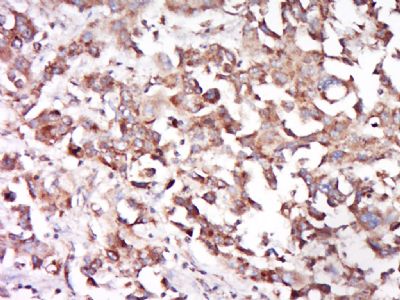
[IF=1.77] Lin, Li-Wen, et al. "Visfatin/pre-B cell colony-enhancing factor immunohistochemical overexpression in oral cancers." Journal of Applied Biomedicine (2014). IHSLCP ; Human.
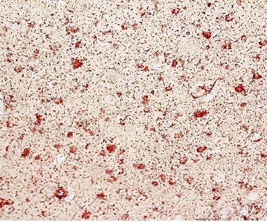
[IF=3.32] Silva, Katie, et al. "Cortical neurons are a prominent source of the proinflammatory cytokine osteopontin in HISLVassociated neurocognitive disorders." Journal of NeuroVirology (2015): 1-12. IHSLCP ; Human.
[IF=3.99] Hu, Kaili, et al. "Lactoferrin conjugated PEG-PLGA nanoparticles for brain delivery: Preparation, characterization and efficacy in Parkinson's disease." International journal of pharmaceutics 415.1 (2011): 273-283. IHSLCP ; Mouse.
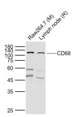
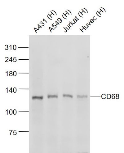
[IF=3.47] Ding L et al. Saponins of sea cucumber attenuate atherosclerosis in ApoE −/− mice via lipid-lowering and anti-inflammatory properties. Journal of Functional Foods,2018 48, 490–497. WB ; Rat&Mouse.
[IF=3.514] Zhang J et al. Role of selective blocking of bradykinin B1 receptor in attenuating immune liver injury in trichloroethylene-sensitized mice. Cytokine. 2018 Aug;108:71-81. IHSLCP ; Mouse.