Home > Product > Antibody > Rabbit Anti-CD80 antibody
Activation B7-1 antigen; B lymphocyte activation antigen B7; B7; B7-1; B7-1 antigen; BB1; CD28 antigen ligand 1; CD28LG; CD28LG1; CD80 antigen (CD28 antigen ligand 1, B7-1 antigen); CD80 antigen; CD80 molecule; CD80_HUMAN; Costimulatory facto
Cat:
SL1479R
Species Reactivity:
Human,Mouse,Rat,
Immunogen:
KLH conjugated synthetic peptide derived from human CD80:111-265/288<Extracellular>
Format:
Liquid
Storage instructions:
Shipped at 4℃. Store at -20 °C for one year. Avoid repeated freeze/thaw cycles.
Concentration:
1mg/ml
Clonality:
Polyclonal
Isotype:
IgG
Applications:
WB=1:500-2000ELISA=1:5000-10000Flow-Cyt=2µg/Testnot yet tested in other applications.optimal dilutions/concentrations should be determined by the end user.
Host:
Rabbit
Product Overview:
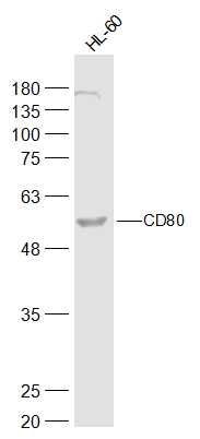
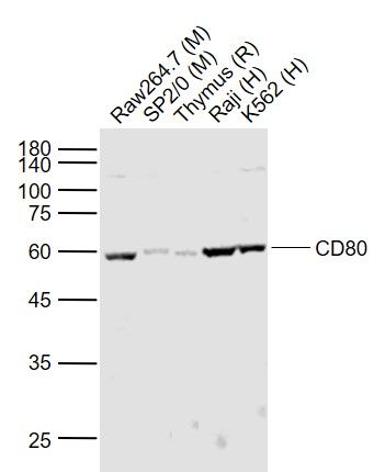
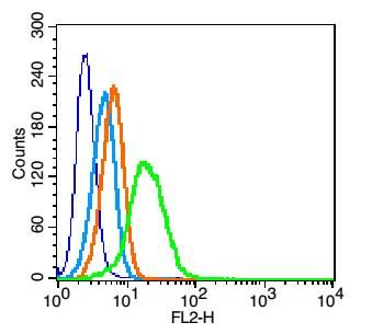
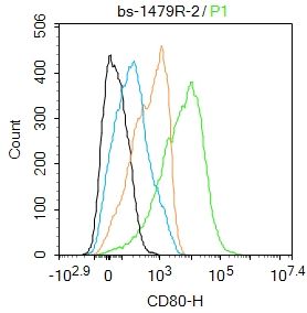
Sample: HL-60(Human) Cell Lysate at 40 ugPrimary: Anti-CD80 (SL1479R) at 1/500 dilutionSecondary: IRDye800CW Goat Anti-Rabbit IgG at 1/20000 dilutionPredicted band size: 32 kDObserved band size: 57 kDSample: Lane 1: Raw264.7 (Mouse) Cell Lysate at 30 ugLane 2: SP2/0 (Mouse) Cell Lysate at 30 ugLane 3: Thymus (Rat) Lysate at 40 ugLane 4: Raji (Human) Cell Lysate at 30 ugLane 5: K562 (Human) Cell Lysate at 30 ugPrimary: Anti-CD80 (SL1479R) at 1/1000 dilutionSecondary: IRDye800CW Goat Anti-Rabbit IgG at 1/20000 dilutionPredicted band size: 60-65 kDObserved band size: 60 kDBlank control: U937(blue). Primary Antibody: Rabbit Anti-CD80 antibody(SL1479R), Dilution: 1μg in 100 μL 1X PBS containing 0.5% BSA; Isotype Control Antibody: Rabbit IgG (orange) ,used under the same conditions. Secondary Antibody: Goat anti-rabbit IgG-PE(white blue), Dilution: 1:200 in 1 X PBS containing 0.5% BSA.ProtocolThe cells were fixed with 2% paraformaldehyde (10 min).Primary antibody (SL1479R, 1μg /1x10^6 cells) were incubated for 30 min on the ice, followed by 1 X PBS containing 0.5% BSA + 10% goat serum (15 min) to block non-specific protein-protein interactions. Then the Goat Anti-rabbit IgG/PE antibody was added into the blocking buffer mentioned above to react with the primary antibody at 1/200 dilution for 30 min on ice. Acquisition of 20,000 events was performed.Blank control:Raji. Primary Antibody (green line): Rabbit Anti-CD80 antibody (SL1479R) Dilution: 2ug/Test; Secondary Antibody : Goat anti-rabbit IgG-AF488Dilution: 0.5ug/Test. ProtocolThe cells were incubated in 5%BSA to block non-specific protein-protein interactions for 30 min at room temperature .Cells stained with Primary Antibody for 30 min at room temperature. The secondary antibody used for 40 min at room temperature. Acquisition of 20,000 events was performed.
More
Unit:
Price: $
Product PDFs
Datasheet:


The protein encoded by this gene is a membrane receptor that is activated by the binding of CD28 or CTLA-4. The activated protein induces T-cell proliferation and cytokine production. This protein can act as a receptor for adenovirus subgroup B and may play a role in lupus neuropathy. [provided by RefSeq, Aug 2011]

Function:
Involved in the costimulatory signal essential for T-lymphocyte activation. T-cell proliferation and cytokine production is induced by the binding of CD28 or CTLA-4 to this receptor.

Subcellular Location:
Membrane.

Tissue Specificity:
Expressed on activated B-cells, macrophages and dendritic cells.

Similarity:
Contains 1 Ig-like C2-type (immunoglobulin-like) domain. Contains 1 Ig-like SLVtype (immunoglobulin-like) domain.

SWISS:
P33681

Gene ID:
941

Database links:

Entrez Gene: 941 Human

Entrez Gene: 12519 Mouse

Omim: 112203 Human

SwissProt: P33681 Human

SwissProt: Q00609 Mouse

Unigene: 838 Human

Unigene: 89474 Mouse


Picture

Sample:
HL-60(Human) Cell Lysate at 40 ug
Primary: Anti-CD80 (SL1479R) at 1/500 dilution
Secondary: IRDye800CW Goat Anti-Rabbit IgG at 1/20000 dilution
Predicted band size: 32 kD
Observed band size: 57 kD
Sample:
Lane 1: Raw264.7 (Mouse) Cell Lysate at 30 ug
Lane 2: SP2/0 (Mouse) Cell Lysate at 30 ug
Lane 3: Thymus (Rat) Lysate at 40 ug
Lane 4: Raji (Human) Cell Lysate at 30 ug
Lane 5: K562 (Human) Cell Lysate at 30 ug
Primary: Anti-CD80 (SL1479R) at 1/1000 dilution
Secondary: IRDye800CW Goat Anti-Rabbit IgG at 1/20000 dilution
Predicted band size: 60-65 kD
Observed band size: 60 kD
Blank control: U937(blue).
Primary Antibody: Rabbit Anti-CD80 antibody(SL1479R), Dilution: 1μg in 100 μL 1X PBS containing 0.5% BSA;
Isotype Control Antibody: Rabbit IgG (orange) ,used under the same conditions.
Secondary Antibody: Goat anti-rabbit IgG-PE(white blue), Dilution: 1:200 in 1 X PBS containing 0.5% BSA.
Protocol
The cells were fixed with 2% paraformaldehyde (10 min).Primary antibody (SL1479R, 1μg /1x10^6 cells) were incubated for 30 min on the ice, followed by 1 X PBS containing 0.5% BSA + 10% goat serum (15 min) to block non-specific protein-protein interactions. Then the Goat Anti-rabbit IgG/PE antibody was added into the blocking buffer mentioned above to react with the primary antibody at 1/200 dilution for 30 min on ice. Acquisition of 20,000 events was performed.
Blank control:Raji.
Primary Antibody (green line): Rabbit Anti-CD80 antibody (SL1479R)
Dilution: 2ug/Test;
Secondary Antibody : Goat anti-rabbit IgG-AF488
Dilution: 0.5ug/Test.
Protocol
The cells were incubated in 5%BSA to block non-specific protein-protein interactions for 30 min at room temperature .Cells stained with Primary Antibody for 30 min at room temperature. The secondary antibody used for 40 min at room temperature. Acquisition of 20,000 events was performed.
Product Feedback Wall
Message :
Your Email :
Copyright © 2007-2018 Sunlong Medical All Rights Reserved.