Home > Product > Antibody > Rabbit Anti-DcR2 antibody
TNFRSF10D; Decoy receptor 2; CD 264; CD264; CD-264; DcR 2; decoy with truncated death domain; ID; TNF receptor related receptor for TRAIL; TNF related apoptosis inducing ligand receptor 4; TNFRSF 10D; TRAIL R4; TRAIL receptor 4; TRAIL receptor with a trun
Cat:
SL1694R
Species Reactivity:
Human,
Immunogen:
KLH conjugated synthetic peptide derived from human DcR2:56-160/386<Extracellular>
Format:
Liquid
Storage instructions:
Shipped at 4℃. Store at -20 °C for one year. Avoid repeated freeze/thaw cycles.
Concentration:
1mg/ml
Clonality:
Polyclonal
Isotype:
IgG
Applications:
WB=1:500-2000ELISA=1:5000-10000Flow-Cyt=1μg /testnot yet tested in other applications.optimal dilutions/concentrations should be determined by the end user.
Host:
Rabbit
Product Overview:
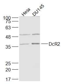
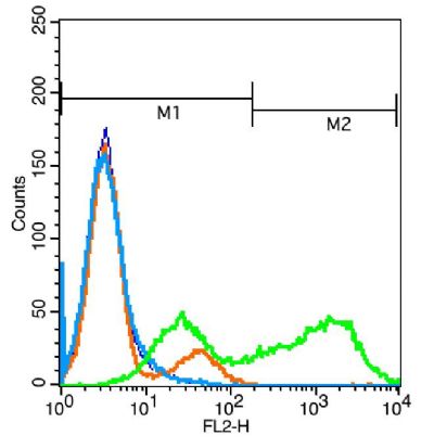
Sample: Hela(Human) Cell Lysate at 30 ugDU145(Human) Cell Lysate at 30 ugPrimary: Anti-DcR2 (SL1694R) at 1/1000 dilutionSecondary: IRDye800CW Goat Anti-Rabbit IgG at 1/20000 dilutionPredicted band size: 36 kDObserved band size: 37 kDBlank control: Jurkat cells(blue). Primary Antibody:Rabbit Anti-DcR2 antibody(SL1694R), Dilution: 1μg in 100 μL 1X PBS containing 0.5% BSA; Isotype Control Antibody: Rabbit IgG(orange) ,used under the same conditions ); Secondary Antibody: Goat anti-rabbit IgG-PE(white blue), Dilution: 1:200 in 1 X PBS containing 0.5% BSA.ProtocolThe cells were fixed with 2% paraformaldehyde (10 min) . Primary antibody (SL1694R, 1μg /1x10^6 cells) were incubated for 30 min on the ice, followed by 1 X PBS containing 0.5% BSA + 1 0% goat serum (15 min) to block non-specific protein-protein interactions. Then the Goat Anti-rabbit IgG/PE antibody was added into the blocking buffer mentioned above to react with the primary antibody at 1/200 dilution for 30 min on ice. Acquisition of 20,000 events was performed.
More
Unit:
Price: $
Product PDFs
Datasheet:


Apoptosis is induced by certain cytokines including TNF and Fas ligand in the TNF family through their death domain containing receptors. TRAIL/Apo2L, a member of the TNF family, induces apoptosis of a variety of tumor cell lines. DR4 and DR5 are functional receptors for TRAIL, and DcR1/TRID is a decoy receptor. Another member of the TRAIL receptor family was identified and designated DcR2. The DcR2 receptor is 386 amino acids in length and has an extracellular TRAIL binding domain, but lacks intracellular death domain and does not induce apoptosis. Although this receptor binds to the cytotoxic ligand TRAIL, it contains a truncated death domain and functions as an inhibitory receptor. When overexpressed, the DcR2 receptor can protect cells against TRAIL mediated cytotoxicity. Like DR4 and DR5, DcR2 transcript is widely expressed in a variety of normal human tissues but DcR2 is absent in most tumors. Ultraviolet radiation has been shown to upregulate DcR2 expression on human keratinocytes. Over expression of DcR2 attenuated TRAIL induced apoptosis.

Function:
Receptor for the cytotoxic ligand TRAIL. Contains a truncated death domain and hence is not capable of inducing apoptosis but protects against TRAIL-mediated apoptosis. Reports are contradictory with regards to its ability to induce the NF-kappa-B pathway. According to PubMed:9382168, it cannot but according to PubMed:9430226, it can induce the NF-kappa-B pathway.

Subcellular Location:
Membrane; Single-pass type I membrane rotein.

Tissue Specificity:
Widely expressed, in particular in fetal kidney, lung and liver, and in adult testis and liver. Also expressed in peripheral blood leukocytes, colon and small intestine, ovary, prostate, thymus, spleen, pancreas, kidney, lung, placenta and heart.

Similarity:
Contains 1 death domain.
Contains 3 TNFR-Cys repeats.

SWISS:
Q9UBN6

Gene ID:
8793

Database links:

Entrez Gene: 8793 Human

Omim: 603614 Human

SwissProt: Q9UBN6 Human

Unigene: 213467 Human


DcR1因缺乏细胞内信号传导系统而不能传递凋亡信号,但能竞争性与TRAIL(肿瘤坏死因子相关凋亡诱导配体)结合,有干扰凋亡信号的传导作用。 Picture

Sample:
Hela(Human) Cell Lysate at 30 ug
DU145(Human) Cell Lysate at 30 ug
Primary: Anti-DcR2 (SL1694R) at 1/1000 dilution
Secondary: IRDye800CW Goat Anti-Rabbit IgG at 1/20000 dilution
Predicted band size: 36 kD
Observed band size: 37 kD
Blank control: Jurkat cells(blue).
Primary Antibody:Rabbit Anti-DcR2 antibody(SL1694R), Dilution: 1μg in 100 μL 1X PBS containing 0.5% BSA;
Isotype Control Antibody: Rabbit IgG(orange) ,used under the same conditions );
Secondary Antibody: Goat anti-rabbit IgG-PE(white blue), Dilution: 1:200 in 1 X PBS containing 0.5% BSA.
Protocol
The cells were fixed with 2% paraformaldehyde (10 min) . Primary antibody (SL1694R, 1μg /1x10^6 cells) were incubated for 30 min on the ice, followed by 1 X PBS containing 0.5% BSA + 1 0% goat serum (15 min) to block non-specific protein-protein interactions. Then the Goat Anti-rabbit IgG/PE antibody was added into the blocking buffer mentioned above to react with the primary antibody at 1/200 dilution for 30 min on ice. Acquisition of 20,000 events was performed.
Product Feedback Wall
Message :
Your Email :
Copyright © 2007-2018 Sunlong Medical All Rights Reserved.