| Picture |
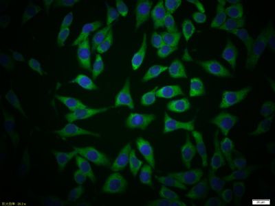
Tissue/cell: MCF-7 cell; 4% Paraformaldehyde-fixed; Triton X-100 at room temperature for 20 min; Blocking buffer (normal goat serum, SLC0005) at 37°C for 20 min; Antibody incubation with (ERG/KCNH2) Polyclonal Antibody, Unconjugated (SL1815R) 1:100, 90 minutes at 37°C; followed by a conjugated Goat Anti-Rabbit IgG antibody (SL0295G-FITC) at 37°C for 90 minutes, DAPI (5ug/ml, blue, SLC0033) was used to stain the cell nuclei.
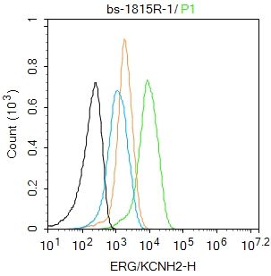
Blank control: RAW264.7.
Primary Antibody (green line): Rabbit Anti-ERG/KCNH2 antibody (SL1815R)
Dilution: 1ug/Test;
Secondary Antibody : Goat anti-rabbit IgG-FITC
Dilution: 0.5ug/Test.
Protocol
The cells were incubated in 5%BSA to block non-specific protein-protein interactions for 30 min at room temperature .Cells stained with Primary Antibody for 30 min at room temperature. The secondary antibody used for 40 min at room temperature. Acquisition of 20,000 events was performed.
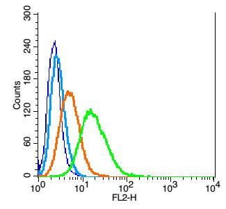
Blank control: RSC96(blue).
Primary Antibody:Rabbit Anti- Cathepsin L antibody(SL1815R), Dilution: 1μg in 100 μL 1X PBS containing 0.5% BSA;
Isotype Control Antibody: Rabbit IgG(orange) ,used under the same conditions );
Secondary Antibody: Goat anti-rabbit IgG-PE(white blue), Dilution: 1:200 in 1 X PBS containing 0.5% BSA.
Protocol
The cells were fixed with 2% paraformaldehyde (10 min). Antibody (SL1815R, 1μg /1x10^6 cells) were incubated for 30 min on the ice, followed by 1 X PBS containing 0.5% BSA + 10% goat serum (15 min) to block non-specific protein-protein interactions. Then the Goat Anti-rabbit IgG/PE antibody was added into the blocking buffer mentioned above to react with the primary antibody of SL1815R at 1/200 dilution for 30 min on ice. Acquisition of 20,000 events was performed.
|