DUSP4 is a member of the dual specificity protein phosphatase subfamily. These phosphatases inactivate their target kinases by dephosphorylating both the phosphoserine/threonine and phosphotyrosine residues. They negatively regulate members of the mitogen-activated protein (MAP) kinase superfamily (MAPK/ERK, SAPK/JNK, p38), which is associated with cellular proliferation and differentiation. Different members of the family of dual specificity phosphatases show distinct substrate specificities for various MAP kinases, different tissue distribution and subcellular localization, and different modes of inducibility of their expression by extracellular stimuli. This gene product inactivates ERK1, ERK2 and JNK, is expressed in a variety of tissues, and is localized in the nucleus. Two alternatively spliced transcript variants, encoding distinct isoforms, have been observed for this gene. In addition, multiple polyadenylation sites have been reported.
Subunit:
Hollow spherical complex composed of 24 subunits with pseudooctahedral symmetry, has a tetramer as the basic unit.
Subcellular Location:
Nucleus.
Similarity:
Belongs to the protein-tyrosine phosphatase family. Non-receptor class dual specificity subfamily.
Contains 1 rhodanese domain.
Contains 1 tyrosine-protein phosphatase domain.
SWISS:
Q13115
Gene ID:
1846
Database links:
Entrez Gene: 1846 Human
Entrez Gene: 319520 Mouse
Omim: 602747 Human
SwissProt: Q13115 Human
SwissProt: Q8SFV3 Mouse
Unigene: 417962 Human
Unigene: 170276 Mouse
Unigene: 392187 Mouse
MKPs是一类丝氨酸/苏氨酸和酪氨酸双重底物特异性的磷酸酶 ,对于丝裂素活化蛋白激酶活性的调节起着十分重要的作用,可使丝裂素活化蛋白激酶上的苏氨酸/酪氨酸去磷酸化失活。目前研究发现MKPs分别有MKP-1、MKP-2、MKP-3及MKP4-6。
MKPs受MAPK信号通路中多种成分的诱导 ,决定了它与MAPK之间作用的特异性。 通过去磷酸化作用调节MAPK信号途径的活性 ,确保了细胞内信号的精确传递 ,参与了多种主要的细胞功能的调节。
| Picture |
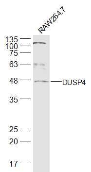
Sample:
RAW264.7(Mouse) Cell Lysate at 30 ug
Primary: Anti-DUSP4 (SL1852R) at 1/500 dilution
Secondary: IRDye800CW Goat Anti-Rabbit IgG at 1/20000 dilution
Predicted band size: 44 kD
Observed band size: 44 kD
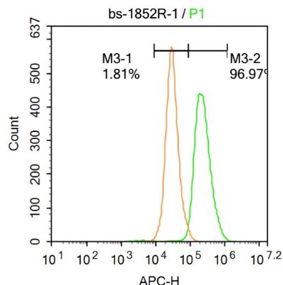
Blank control:A431.
Primary Antibody (green line): Rabbit Anti-DUSP4 antibody (SL1852R)
Dilution: 1μg /10^6 cells;
Isotype Control Antibody (orange line): Rabbit IgG .
Secondary Antibody : Goat anti-rabbit IgG-AF647
Dilution: 1μg /test.
Protocol
The cells were fixed with 4% PFA (10min at room temperature)and then permeabilized with 90% ice-cold methanol for 20 min at-20℃. The cells were then incubated in 5%BSA to block non-specific protein-protein interactions for 30 min at at room temperature .Cells stained with Primary Antibody for 30 min at room temperature. The secondary antibody used for 40 min at room temperature. Acquisition of 20,000 events was performed.
|
|
|