Home > Product > Antibody > Rabbit Anti-SV2A antibody
Synaptic vesicle protein 2A; Synaptic vesicle glycoprotein 2A; Synaptic vesicle protein 2A; Synaptic vesicle protein 2; Sv2; Kiaa0736; SV2A_HUMAN.
Cat:
SL487R
Species Reactivity:
Human,(predicted: Mouse,Rat,Dog,Pig,Cow,Horse,Rabbit,)
Immunogen:
KLH conjugated synthetic peptide derived from human SV2A:451-550/743<Extracellular>
Format:
Liquid
Storage instructions:
Shipped at 4℃. Store at -20 °C for one year. Avoid repeated freeze/thaw cycles.
Concentration:
1mg/ml
Clonality:
Polyclonal
Isotype:
IgG
Applications:
ELISA=1:5000-10000Flow-Cyt=1ug/testnot yet tested in other applications.optimal dilutions/concentrations should be determined by the end user.
Host:
Rabbit
Product Overview:
Black line : Positive blank control (Molt4); Negative blank control (A549) Green line : Primary Antibody (Rabbit Anti- SV2A antibody (SL487R) ) Orange line:Isotype Control Antibody (Rabbit IgG) . Blue line : Secondary Antibody (Goat anti-rabbit IgG-AF488)/(Goat anti-rabbit IgG-AF647) Molt4(Positive)and A549(Negative control)cells (black) were fixed with 4% PFA for 10min at room temperature, permeabilized with PBST for 20 min at room temperature, and incubated in 5% BSA blocking buffer for 30 min at room temperature. Cells were then stained with SV2A Antibody(SL487R)at 1:50 dilution in blocking buffer and incubated for 30 min at room temperature, washed twice with 2% BSA in PBS, followed by secondary antibody(blue) incubation for 40 min at room temperature. Acquisitions of 20,000 events were performed. Cells stained with primary antibody (green), and isotype control (orange).Blank control:Molt-4. Primary Antibody (green line): Rabbit Anti-SV2A antibody (SL487) Dilution: 1μg /10^6 cells; Isotype Control Antibody (orange line): Rabbit IgG . Secondary Antibody : Goat anti-rabbit IgG-PEDilution: 1μg /test. ProtocolThe cells were fixed with 4% PFA (10min at room temperature)and then permeabilized with 20% PBST for 20 min at-20℃. The cells were then incubated in 5%BSA to block non-specific protein-protein interactions for 30 min at at room temperature .Cells stained with Primary Antibody for 30 min at room temperature. The secondary antibody used for 40 min at room temperature. Acquisition of 20,000 events was performed.
More
Unit:
Price: $
Product PDFs
Datasheet:


SV2s (Synaptic Vesicle protein 2) are integral membrane glycoproteins present in all synaptic vesicles. They have 12 transmembrane domains predicted by sequence analysis. There are three characterized isoforms, SV2A, SV2B and SV2C. SV2A is expressed ubiquitously throughout the brain. SV2B has a more restricted distribution with varying degrees of coexpression with SV2A. SV2C is more closely related to SV2A but shows a very restricted expression pattern. The highest expression levels were observed in phylogenetically old brain areas like pallidum, the midbrain and the olfactory bulb. SV2A is probably involved in the maintenance of a pool of synaptic vesicles competent for calcium-stimulated exocytosis. SV2A is a highly glycosylated synaptic vesicle protein with homology to transmembrane transporters.

Function:
Plays a role in the control of regulated secretion in neural and endocrine cells, enhancing selectively low-frequency neurotransmission. Positively regulates vesicle fusion by maintaining the readily releasable pool of secretory vesicles (By similarity).

Subunit:
Interacts with SYT1/synaptotagmin-1 in a calcium-dependent manner. Binds the adapter protein complex AP-2.

Subcellular Location:
Cytoplasmic vesicle, secretory vesicle, synaptic vesicle membrane; Multi-pass membrane protein.

Post-translational modifications:
Phosphorylation by CK1 of the N-terminal cytoplasmic domain regulates interaction with SYT1.
N-glycosylated.

Similarity:
Belongs to the major facilitator superfamily.

SWISS:
Q7L0J3

Gene ID:
9900

Database links:

Entrez Gene: 9900 Human

Entrez Gene: 12851 Mouse

Entrez Gene: 117559 Rat

Omim: 185860 Human

SwissProt: Q29397 Cow

SwissProt: Q7L0J3 Human

SwissProt: Q9JIS5 Mouse

SwissProt: Q02563 Rat

Unigene: 516153 Human

Unigene: 200365 Mouse

Unigene: 11264 Rat


突触泡蛋白2A(SV2A)是一类跨膜糖蛋白,定位于脊椎动物神经元及内分泌细胞,与神经递质的释放、内分泌泡胞吐作用、突触泡稳态的维持作用。 Picture

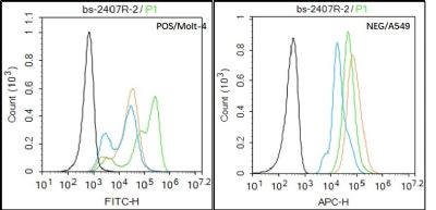
Black line : Positive blank control (Molt4); Negative blank control (A549)
Green line : Primary Antibody (Rabbit Anti- SV2A antibody (SL487R) )
Orange line:Isotype Control Antibody (Rabbit IgG) .
Blue line : Secondary Antibody (Goat anti-rabbit IgG-AF488)/(Goat anti-rabbit IgG-AF647)
Molt4(Positive)and A549(Negative control)cells (black) were fixed with 4% PFA for 10min at room temperature, permeabilized with PBST for 20 min at room temperature, and incubated in 5% BSA blocking buffer for 30 min at room temperature. Cells were then stained with SV2A Antibody(SL487R)at 1:50 dilution in blocking buffer and incubated for 30 min at room temperature, washed twice with 2% BSA in PBS, followed by secondary antibody(blue) incubation for 40 min at room temperature. Acquisitions of 20,000 events were performed. Cells stained with primary antibody (green), and isotype control (orange).
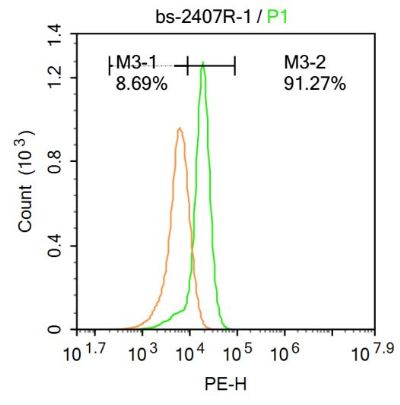
Blank control:Molt-4.
Primary Antibody (green line): Rabbit Anti-SV2A antibody (SL487)
Dilution: 1μg /10^6 cells;
Isotype Control Antibody (orange line): Rabbit IgG .
Secondary Antibody : Goat anti-rabbit IgG-PE
Dilution: 1μg /test.
Protocol
The cells were fixed with 4% PFA (10min at room temperature)and then permeabilized with 20% PBST for 20 min at-20℃. The cells were then incubated in 5%BSA to block non-specific protein-protein interactions for 30 min at at room temperature .Cells stained with Primary Antibody for 30 min at room temperature. The secondary antibody used for 40 min at room temperature. Acquisition of 20,000 events was performed.
Product Feedback Wall
Message :
Your Email :
Copyright © 2007-2018 Sunlong Medical All Rights Reserved.