Home > Product > Antibody > Rabbit Anti-SDHA antibody
mitochondrial; DHSA_HUMAN; Flavoprotein subunit of complex II; Fp; SDH 2; SDH2; SDHA; SDHF; Succinate dehydrogenase [ubiquinone] flavoprotein subunit; Succinate dehydrogenase [ubiquinone] flavoprotein subunit mitochondrial; Succinate dehydrogenase complex
Cat:
SL3970R
Species Reactivity:
Human,Mouse,(predicted: Rat,)
Immunogen:
KLH conjugated synthetic peptide derived from human SDHA:561-664/664
Format:
Liquid
Storage instructions:
Shipped at 4℃. Store at -20 °C for one year. Avoid repeated freeze/thaw cycles.
Concentration:
1mg/ml
Clonality:
Polyclonal
Isotype:
IgG
Applications:
WB=1:500-2000ELISA=1:5000-10000IHC-P=1:100-500IHC-F=1:100-500Flow-Cyt=2ug/TestIF=1:100-500(Paraffin sections need to do antigen repair)not yet tested in other applications.optimal dilutions/concentrations should be determined by the end user.
Host:
Rabbit
Product Overview:
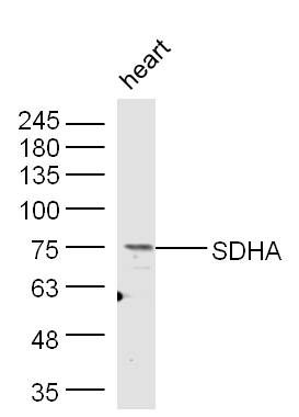
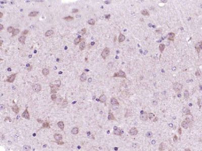
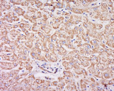
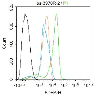
Sample: Heart (Mouse) Lysate at 40 ugPrimary: Anti- SDHA (SL3970R) at 1/300 dilutionSecondary: IRDye800CW Goat Anti-Rabbit IgG at 1/10000 dilutionPredicted band size: 70 kDObserved band size: 75 kDParaformaldehyde-fixed, paraffin embedded (mouse brain); Antigen retrieval by boiling in sodium citrate buffer (pH6.0) for 15min; Block endogenous peroxidase by 3% hydrogen peroxide for 20 minutes; Blocking buffer (normal goat serum) at 37°C for 30min; Antibody incubation with (SDHA) Polyclonal Antibody, Unconjugated (SL3970R) at 1:200 overnight at 4°C, followed by operating according to SP Kit(Rabbit) (sp-0023) instructionsand DAB staining.Tissue/cell: human liver cancer; 4% Paraformaldehyde-fixed and paraffin-embedded; Antigen retrieval: citrate buffer ( 0.01M, pH 6.0 ), Boiling bathing for 15min; Block endogenous peroxidase by 3% Hydrogen peroxide for 30min; Blocking buffer (normal goat serum,SLC0005) at 37℃ for 20 min; Incubation: Anti-SDHA Polyclonal Antibody, Unconjugated(SL3970R) 1:200, overnight at 4°C, followed by conjugation to the secondary antibody(SP-0023) and DAB(SLC0010) stainingBlank control: RAW264.7. Primary Antibody (green line): Rabbit Anti-SDHA antibody (SL3970R) Dilution: 2ug/Test; Secondary Antibody : Goat anti-rabbit IgG-FITCDilution: 0.5ug/Test. ProtocolThe cells were fixed with 4% PFA (10min at room temperature)and then permeabilized with 0.1% PBST for 20 min at room temperature.The cells were then incubated in 5%BSA to block non-specific protein-protein interactions for 30 min at room temperature .Cells stained with Primary Antibody for 30 min at room temperature. The secondary antibody used for 40 min at room temperature. Acquisition of 20,000 events was performed.
More
Unit:
Price: $
Product PDFs
Datasheet:


In aerobic respiration reactions, succinate dehydrogenase (SDH) catalyzes the oxidation of succinate and ubiquinone to fumarate and ubiquinol. Four subunits comprise the SDH protein complex: a flavochrome subunit (SDHA), an iron-sulfur protein (SDHB), and two membrane-bound subunits (SDHC and SDHD) anchored to the inner mitochondrial membrane. Mutations to these subunits cause mitochondrial dysfunction, corresponding to several distinct disorders. Mutations in the membrane bound components may cause hereditary paraganglioma, while SDHA mutations are associated with juvenile encephalopathy as well as Leigh Syndrome, a severe neurological disorder. Inactivating mutations in SDHB correlate with inherited, but not necessarily sporadic, cases of pheochromocytoma.

Function:
Defects in SDHA are a cause of mitochondrial complex II deficiency (MT-C2D). A disorder of the mitochondrial respiratory chain with heterogeneous clinical manifestations. Clinical features include psychomotor regression in infants, poor growth with lack of speech development, severe spastic quadriplegia, dystonia, progressive leukoencephalopathy, muscle weakness, exercise intolerance, cardiomyopathy. Some patients manifest Leigh syndrome or Kearns-Sayre syndrome.

SWISS:
P31040

Gene ID:
6389

Picture

Sample: Heart (Mouse) Lysate at 40 ug
Primary: Anti- SDHA (SL3970R) at 1/300 dilution
Secondary: IRDye800CW Goat Anti-Rabbit IgG at 1/10000 dilution
Predicted band size: 70 kD
Observed band size: 75 kD
Paraformaldehyde-fixed, paraffin embedded (mouse brain); Antigen retrieval by boiling in sodium citrate buffer (pH6.0) for 15min; Block endogenous peroxidase by 3% hydrogen peroxide for 20 minutes; Blocking buffer (normal goat serum) at 37°C for 30min; Antibody incubation with (SDHA) Polyclonal Antibody, Unconjugated (SL3970R) at 1:200 overnight at 4°C, followed by operating according to SP Kit(Rabbit) (sp-0023) instructionsand DAB staining.
Tissue/cell: human liver cancer; 4% Paraformaldehyde-fixed and paraffin-embedded;
Antigen retrieval: citrate buffer ( 0.01M, pH 6.0 ), Boiling bathing for 15min; Block endogenous peroxidase by 3% Hydrogen peroxide for 30min; Blocking buffer (normal goat serum,SLC0005) at 37℃ for 20 min;
Incubation: Anti-SDHA Polyclonal Antibody, Unconjugated(SL3970R) 1:200, overnight at 4°C, followed by conjugation to the secondary antibody(SP-0023) and DAB(SLC0010) staining
Blank control: RAW264.7.
Primary Antibody (green line): Rabbit Anti-SDHA antibody (SL3970R)
Dilution: 2ug/Test;
Secondary Antibody : Goat anti-rabbit IgG-FITC
Dilution: 0.5ug/Test.
Protocol
The cells were fixed with 4% PFA (10min at room temperature)and then permeabilized with 0.1% PBST for 20 min at room temperature.The cells were then incubated in 5%BSA to block non-specific protein-protein interactions for 30 min at room temperature .Cells stained with Primary Antibody for 30 min at room temperature. The secondary antibody used for 40 min at room temperature. Acquisition of 20,000 events was performed.
Product Feedback Wall
Message :
Your Email :
Copyright © 2007-2018 Sunlong Medical All Rights Reserved.