NCF1, along with NCF2 and a membrane bound cytochrome b558, is required for activation of the latent NADPH oxidase necessary for superoxide production. Defects in NCF1 are the cause of autosomal cytochrome-b-positive chronic granulomatous disease type 1 (CGD).
Function:
NCF2, NCF1, and a membrane bound cytochrome b558 are required for activation of the latent NADPH oxidase (necessary for superoxide production).
Subunit:
Interacts with NOXA1. Interacts with ADAM15. Interacts with TRAF4. Interacts with FASLG.
Subcellular Location:
Cytoplasm.
Post-translational modifications:
Phosphorylated by PRKCD; phosphorylation induces activation of NCF1 and NADPH oxidase activity.
DISEASE:
Granulomatous disease, chronic, cytochrome-b-positive 1, autosomal recessive (CGD1) [MIM:233700]: A disorder characterized by the inability of neutrophils and phagocytes to kill microbes that they have ingested. Patients suffer from life-threatening bacterial/fungal infections. Note=The disease is caused by mutations affecting the gene represented in this entry.
Similarity:
Contains 1 PX (phox homology) domain.
Contains 2 SH3 domains.
SWISS:
P14598
Gene ID:
653361
Database links:
Entrez Gene: 281345 Cow
Entrez Gene: 653361 Human
Entrez Gene: 17969 Mouse
Entrez Gene: 100134857 Pig
Entrez Gene: 100001763 Rabbit
Entrez Gene: 114553 Rat
Omim: 608512 Human
SwissProt: O77774 Cow
SwissProt: P14598 Human
SwissProt: Q09014 Mouse
Unigene: 647047 Human
Unigene: 655201 Human
Unigene: 425296 Mouse
Unigene: 38575 Rat
| Picture |
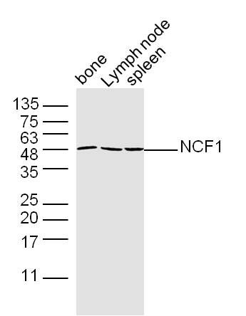
Sample:
Bone (Mouse) Lysate at 40 ug
Lymph node (Mouse) Lysate at 40 ug
Spleen (Mouse) Lysate at 40 ug
Primary: Anti-NCF1 (SL3886R) at 1/300 dilution
Secondary: IRDye800CW Goat Anti-Rabbit IgG at 1/20000 dilution
Predicted band size: 45 kD
Observed band size: 48 kD
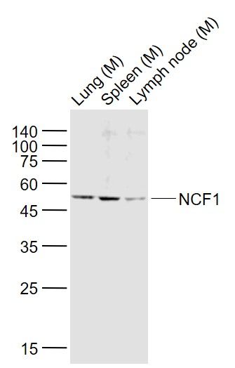
Sample:
Lane 1: Lung (Mouse) Lysate at 40 ug
Lane 2: Spleen (Mouse) Lysate at 40 ug
Lane 3: Lymph node (Mouse) Lysate at 40 ug
Primary: Anti-NCF1 (SL3886R) at 1/1000 dilution
Secondary: IRDye800CW Goat Anti-Rabbit IgG at 1/20000 dilution
Predicted band size: 48 kD
Observed band size: 48 kD
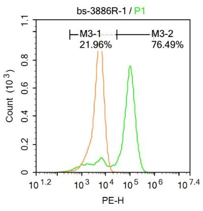
U-937 cells were fixed with 4% PFA for 10min at room temperature,permeabilized with 90% ice-cold methanol for 20 min at room temperature, and incubated in 5% BSA blocking buffer for 30 min at room temperature. Cells were then stained with NCF1 Antibody(SL3886R)at 1:100 dilution in blocking buffer and incubated for 30 min at room temperature, washed twice with 2%BSA in PBS, followed by secondary antibody incubation for 40 min at room temperature. Acquisitions of 20,000 events were performed. Cells stained with primary antibody (green), and isotype control (orange).
|
|
|