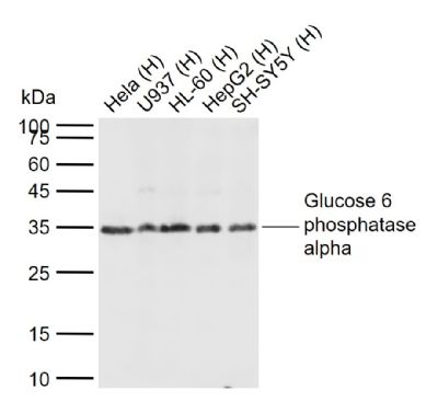
Sample:
Lane 1: Human Hela cell lysates
Lane 2: Human U937cell lysates
Lane 3: Human HL-60 cell lysates
Lane 4: Human HepG2 cell lysates
Lane 5: Human SH-SY5Y cell lysates
Primary: Anti-Glucose 6 phosphatase alpha (SL4044R) at 1/1000 dilution
Secondary: IRDye800CW Goat Anti-Rabbit IgG at 1/20000 dilution
Predicted band size: 39 kDa
Observed band size: 35 kDa
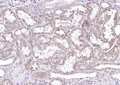
Paraformaldehyde-fixed, paraffin embedded (Human kidney); Antigen retrieval by boiling in sodium citrate buffer (pH6.0) for 15min; Block endogenous peroxidase by 3% hydrogen peroxide for 20 minutes; Blocking buffer (normal goat serum) at 37°C for 30min; Antibody incubation with (Glucose 6 phosphatase alpha) Polyclonal Antibody, Unconjugated (SL4044R) at 1:200 overnight at 4°C, followed by operating according to SP Kit(Rabbit) (sp-0023) instructionsand DAB staining.
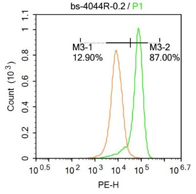
U-937 cells were incubated in 5% BSA blocking buffer for 30 min at room temperature. Cells were then stained with SL4044R Antibody at 1:500 dilution in blocking buffer and incubated for 30 min at room temperature, washed twice with 2%BSA in PBS, followed by secondary antibody incubation for 40 min at room temperature. Acquisitions of 20,000 events were performed. Cells stained with primary antibody (green), and isotype control (orange).
U-937 cells were incubated in 5% BSA blocking buffer for 30 min at room temperature. Cells were then stained with SL4044R Antibody at 1:500 dilution in blocking buffer and incubated for 30 min at room temperature, washed twice with 2%BSA in PBS, followed by secondary antibody incubation for 40 min at room temperature. Acquisitions of 20,000 events were performed. Cells stained with primary antibody (green), and isotype control (orange).
|