Home > Product > Antibody > Rabbit Anti-DP1 antibody
DRTF1; DRTF 1; DRTF-1; TFDP1; TFDP 1; TFDP-1; Transcription factor Dp-1; DRTF1-polypeptide 1; E2F dimerization partner 1; TFDP1_HUMAN.
Cat:
SL5143R
Species Reactivity:
Human,Mouse,Rat,(predicted: Chicken,Dog,Cow,Horse,Rabbit,)
Immunogen:
KLH conjugated synthetic peptide derived from human TFDP1:221-320/410
Format:
Liquid
Storage instructions:
Shipped at 4℃. Store at -20 °C for one year. Avoid repeated freeze/thaw cycles.
Concentration:
1mg/ml
Clonality:
Polyclonal
Isotype:
IgG
Applications:
WB=1:500-2000ELISA=1:5000-10000IHC-P=1:100-500IHC-F=1:100-500IF=1:100-500(Paraffin sections need to do antigen repair)not yet tested in other applications.optimal dilutions/concentrations should be determined by the end user.
Host:
Rabbit
Product Overview:
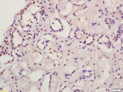
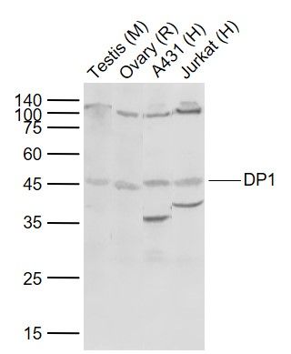
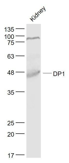
Sample: Lane 1: Mouse Testis tissue lysates Lane 2: Rat Ovary tissue lysates Lane 3: Human A431 cell lysates Lane 4: Human Jurkat cell lysates Primary: Anti-DP1 (SL5143R) at 1/1000 dilutionSecondary: IRDye800CW Goat Anti-Rabbit IgG at 1/20000 dilutionPredicted band size: 45 kDObserved band size: 45 kDSample: Kidney (Mouse) Lysate at 40 ugPrimary: Anti- DP1 (SL5143R) at 1/1000 dilutionSecondary: IRDye800CW Goat Anti-Rabbit IgG at 1/20000 dilutionPredicted band size: 45 kDObserved band size: 45 kDTissue/cell: human kidney tissue; 4% Paraformaldehyde-fixed and paraffin-embedded; Antigen retrieval: citrate buffer ( 0.01M, pH 6.0 ), Boiling bathing for 15min; Block endogenous peroxidase by 3% Hydrogen peroxide for 30min; Blocking buffer (normal goat serum,SLC0005) at 37℃ for 20 min; Incubation: Anti-DP1/TFDP1 Polyclonal Antibody, Unconjugated(SL5143R) 1:200, overnight at 4°C, followed by conjugation to the secondary antibody(SP-0023) and DAB(SLC0010) staining
More
Unit:
Price: $
Product PDFs
Datasheet:


This gene encodes a member of a family of transcription factors that heterodimerize with E2F proteins to enhance their DNA-binding activity and promote transcription from E2F target genes. The encoded protein functions as part of this complex to control the transcriptional activity of numerous genes involved in cell cycle progression from G1 to S phase. Alternative splicing results in multiple transcript variants. Pseudogenes of this gene are found on chromosomes 1, 15, and X.[provided by RefSeq, Jan 2009]

Function:
The DP2/E2F complex functions in the control of cell-cycle progression from G1 to S phase. The E2F-1/DP complex appears to mediate both cell proliferation and apoptosis.

Subunit:
Component of the E2F/DP transcription factor complex. Forms heterodimers with E2F family members. The complex can interact with hypophosphorylated retinoblastoma protein RB1 and related proteins (RBL1 and RBL2) that inhibit the E2F transactivation domain. This repression involves recruitment of histone deacetylase (HDAC). During the cell cycle, from mid-to-late G1 phase, RB family members become phosphorylated, detach from the DRTF1/E2F complex to render E2F transcriptionally active. Viral oncoproteins, notably E1A, T-antigen and HPV E7, are capable of sequestering RB protein, thus releasing the active complex. Part of the E2F6.com-1 complex in G0 phase is composed of E2F6, MGA, MAX, TFDP1, CBX3, BAT8, EUHMTASE1, RING1, RNF2, MBLR, L3MBTL2 YAF2. Component of the DREAM complex (also named LINC complex) at least composed of E2F4, E2F5, LIN9, LIN37, LIN52, LIN54, MYBL1, MYBL2, RBL1, RBL2, RBBP4, TFDP1 and TFDP2. The complex exists in quiescent cells where it represses cell cycle-dependent genes. It dissociates in S phase when LIN9, LIN37, LIN52 and LIN54 form a subcomplex that binds to MYBL2.

Subcellular Location:
Nucleus.

Tissue Specificity:
Highest levels in muscle. Also expressed in brain, placenta, liver and kidney. Lower levels in lung and pancreas.

Post-translational modifications:
Phosphorylation by E2F-1-bound cyclin A-CDK2, in the S phase, inhibits E2F-mediated DNA binding and transactivation.

Similarity:
Belongs to the E2F/DP family.

SWISS:
Q14186

Gene ID:
7027

Database links:

Entrez Gene: 7027 Human

Entrez Gene: 21781 Mouse

Entrez Gene: 290878 Rat

Omim: 189902 Human

SwissProt: Q14186 Human

SwissProt: Q5JSB4 Human

SwissProt: Q08639 Mouse

SwissProt: Q3V3X3 Mouse

SwissProt: Q9CYZ7 Mouse

SwissProt: Q9D297 Mouse

Unigene: 79353 Human

Unigene: 925 Mouse


Picture

Sample:
Lane 1: Mouse Testis tissue lysates
Lane 2: Rat Ovary tissue lysates
Lane 3: Human A431 cell lysates
Lane 4: Human Jurkat cell lysates
Primary: Anti-DP1 (SL5143R) at 1/1000 dilution
Secondary: IRDye800CW Goat Anti-Rabbit IgG at 1/20000 dilution
Predicted band size: 45 kD
Observed band size: 45 kD
Sample:
Kidney (Mouse) Lysate at 40 ug
Primary: Anti- DP1 (SL5143R) at 1/1000 dilution
Secondary: IRDye800CW Goat Anti-Rabbit IgG at 1/20000 dilution
Predicted band size: 45 kD
Observed band size: 45 kD
Tissue/cell: human kidney tissue; 4% Paraformaldehyde-fixed and paraffin-embedded;
Antigen retrieval: citrate buffer ( 0.01M, pH 6.0 ), Boiling bathing for 15min; Block endogenous peroxidase by 3% Hydrogen peroxide for 30min; Blocking buffer (normal goat serum,SLC0005) at 37℃ for 20 min;
Incubation: Anti-DP1/TFDP1 Polyclonal Antibody, Unconjugated(SL5143R) 1:200, overnight at 4°C, followed by conjugation to the secondary antibody(SP-0023) and DAB(SLC0010) staining
Product Feedback Wall
Message :
Your Email :
Copyright © 2007-2018 Sunlong Medical All Rights Reserved.