SNF5 is involved in transcriptional activation. The SWI/SNF complex is required for the induced expression of a large number of genes. This complex alters chromatin structure to facilitate binding of gene-specific dedicated transcription factors. SNF5 also funtions as a tumor suppressor gene and is frequently mutated in malignant rhabdoid tumors. It is also involved in adipocyte differentiation.
Function:
Core component of the BAF (hSWI/SNF) complex. This ATP-dependent chromatin-remodeling complex plays important roles in cell proliferation and differentiation, in cellular antiviral activities and inhibition of tumor formation. The BAF complex is able to create a stable, altered form of chromatin that constrains fewer negative supercoils than normal. This change in supercoiling would be due to the conversion of up to one-half of the nucleosomes on polynucleosomal arrays into asymmetric structures, termed altosomes, each composed of 2 histones octamers. Stimulates in vitro the remodeling activity of SMARCA4/BRG1/BAF190A. Involved in activation of CSF1 promoter. Belongs to the neural progenitors-specific chromatin remodeling complex (npBAF complex) and the neuron-specific chromatin remodeling complex (nBAF complex). During neural development a switch from a stem/progenitor to a post-mitotic chromatin remodeling mechanism occurs as neurons exit the cell cycle and become committed to their adult state. The transition from proliferating neural stem/progenitor cells to post-mitotic neurons requires a switch in subunit composition of the npBAF and nBAF complexes. As neural progenitors exit mitosis and differentiate into neurons, npBAF complexes which contain ACTL6A/BAF53A and PHF10/BAF45A, are exchanged for homologous alternative ACTL6B/BAF53B and DPF1/BAF45B or DPF3/BAF45C subunits in neuron-specific complexes (nBAF). The npBAF complex is essential for the self-renewal/proliferative capacity of the multipotent neural stem cells. The nBAF complex along with CREST plays a role regulating the activity of genes essential for dendrite growth (By similarity). Plays a key role in cell-cycle control and causes cell cycle arrest in G0/G1. Also involved in vitamin D-coupled transcription regulation via its association with the WINAC complex, a chromatin-remodeling complex recruited by vitamin D receptor (VDR), which is required for the ligand-bound VDR-mediated transrepression of the CYP27B1 gene.
Subunit:
Component of the BAF (hSWI/SNF) complex, which includes at least actin (ACTB), ARID1A, ARID1B/BAF250, SMARCA2, SMARCA4/BRG1/BAF190A, ACTL6A/BAF53, ACTL6B/BAF53B, SMARCE1/BAF57 SMARCC1/BAF155, SMARCC2/BAF170, SMARCB1/SNF5/INI1, and one or more of SMARCD1/BAF60A, SMARCD2/BAF60B, or SMARCD3/BAF60C. In muscle cells, the BAF complex also contains DPF3. Component of the WINAC complex, at least composed of SMARCA2, SMARCA4, SMARCB1, SMARCC1, SMARCC2, SMARCD1, SMARCE1, ACTL6A, BAZ1B/WSTF, ARID1A, SUPT16H, CHAF1A and TOP2B. Binds to double-stranded DNA. Interacts with MYK and MAEL. Interacts with PPP1R15A. Binds tightly to the human immunodeficiency virus-type 1 (HISLV1) integrase in vitro and stimulates its DNA-joining activity. Interacts with human papillomavirus 18 E1 protein to stimulate its viral replication. Interacts with Epstein-Barr virus protein EBNA-2. Component of neural progenitors-specific chromatin remodeling complex (npBAF complex) composed of at least, ARID1A/BAF250A or ARID1B/BAF250B, SMARCD1/BAF60A, SMARCD3/BAF60C, SMARCA2/BRM/BAF190B, SMARCA4/BRG1/BAF190A, SMARCB1/BAF47, SMARCC1/BAF155, SMARCE1/BAF57, SMARCC2/BAF170, PHF10/BAF45A, ACTL6A/BAF53A and actin. Component of neuron-specific chromatin remodeling complex (nBAF complex) composed of at least, ARID1A/BAF250A or ARID1B/BAF250B, SMARCD1/BAF60A, SMARCD3/BAF60C, SMARCA2/BRM/BAF190B, SMARCA4/BRG1/BAF190A, SMARCB1/BAF47, SMARCC1/BAF155, SMARCE1/BAF57, SMARCC2/BAF170, DPF1/BAF45B, DPF3/BAF45C, ACTL6B/BAF53B and actin (By similarity).
Subcellular Location:
Nucleus.
Post-translational modifications:
Phosphorylated upon DNA damage, probably by ATM or ATR.
DISEASE:
Defects in SMARCB1 are a cause of schwannomatosis (SCHWA) [MIM:162091]; also called congenital cutaneous neurilemmomatosis. Schwannomas are benign tumors of the peripheral nerve sheath that usually occur singly in otherwise normal individuals. Multiple schwannomas in the same individual suggest an underlying tumor-predisposition syndrome. The most common such syndrome is NF2. The hallmark of NF2 is the development of bilateral vestibular-nerve schwannomas; but two-thirds or more of all NF2-affected individuals develop schwannomas in other locations, and dermal schwannomas may precede vestibular tumors in NF2-affected children. There have been several reports of individuals with multiple schwannomas who do not show evidence of vestibular schwannoma. Clinical report suggests that schwannomatosis is a clinical entity distinct from other forms of neurofibromatosis.
Similarity:
Belongs to the SNF5 family.
SWISS:
Q12824
Gene ID:
6598
Database links:
Entrez Gene: 537412 Cow
Entrez Gene: 481287 Dog
Entrez Gene: 6598 Human
Entrez Gene: 20587 Mouse
Entrez Gene: 100154735 Pig
Entrez Gene: 361825 Rat
Entrez Gene: 443934 Xenopus laevis
Omim: 601607 Human
SwissProt: Q5BIN2 Cow
SwissProt: Q12824 Human
SwissProt: Q9Z0H3 Mouse
SwissProt: Q6GQ82 Xenopus laevis
Unigene: 534350 Human
Unigene: 279751 Mouse
Unigene: 405473 Mouse
Unigene: 44271 Xenopus laevis
| Picture |
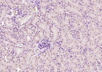
Paraformaldehyde-fixed, paraffin embedded (mouse kidney); Antigen retrieval by boiling in sodium citrate buffer (pH6.0) for 15min; Block endogenous peroxidase by 3% hydrogen peroxide for 20 minutes; Blocking buffer (normal goat serum) at 37°C for 30min; Antibody incubation with (SNF5) Polyclonal Antibody, Unconjugated (SL6109R) at 1:200 overnight at 4°C, followed by operating according to SP Kit(Rabbit) (sp-0023) instructionsand DAB staining.
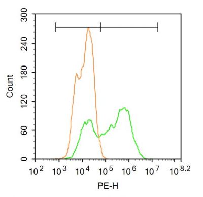
Blank control:A549.
Primary Antibody (green line): Rabbit Anti-SNF5 antibody (SL6109R)
Dilution: 1μg /10^6 cells;
Isotype Control Antibody (orange line): Rabbit IgG .
Secondary Antibody : Goat anti-rabbit IgG-PE
Dilution: 3μg /test.
Protocol
The cells were fixed with 4% PFA (10min at room temperature)and then permeabilized with 90% ice-cold methanol for 20 min at-20℃. The cells were then incubated in 5% BSA to block non-specific protein-protein interactions for 30 min at at room temperature .Cells stained with Primary Antibody for 30 min at room temperature. The secondary antibody used for 40 min at room temperature. Acquisition of 20,000 events was performed.
|
|
|