Home > Product > Antibody > Rabbit Anti-BST1 antibody
Cyclic ADP ribose hydrolase 2; ADP ribosyl cyclase 2; Bone marrow stromal antigen 1; Bone marrow stromal cell antigen 1; BST 1; BST1; BST-1; cADPr hydrolase 2; CD157; CD157 antigen; NAD(+) nucleosidase; BST1_HUMAN.
Cat:
SL6023R
Species Reactivity:
Mouse,(predicted: Human,Rat,Dog,Pig,Cow,Rabbit,)
Immunogen:
KLH conjugated synthetic peptide derived from human BST1/CD157:51-150/318
Format:
Liquid
Storage instructions:
Shipped at 4℃. Store at -20 °C for one year. Avoid repeated freeze/thaw cycles.
Concentration:
1mg/ml
Clonality:
Polyclonal
Isotype:
IgG
Applications:
WB=1:500-2000ELISA=1:5000-10000Flow-Cyt=1μg/Testnot yet tested in other applications.optimal dilutions/concentrations should be determined by the end user.
Host:
Rabbit
Product Overview:
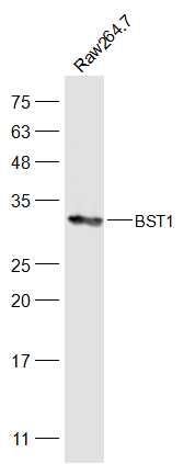
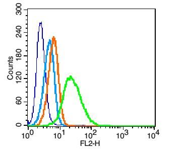
Sample: Raw264.7(Mouse) Cell Lysate at 30 ugPrimary: Anti-BST1 (SL6023R) at 1/500 dilutionSecondary: IRDye800CW Goat Anti-Rabbit IgG at 1/20000 dilutionPredicted band size: 33 kDObserved band size: 33 kDBlank control: U937(blue). Primary Antibody: Rabbit Anti-BST1 antibody(SL6023R), Dilution: 1μg in 100 μL 1X PBS containing 0.5% BSA; Isotype Control Antibody: Rabbit IgG (orange) ,used under the same conditions. Secondary Antibody: Goat anti-rabbit IgG-PE(white blue), Dilution: 1:200 in 1 X PBS containing 0.5% BSA.ProtocolThe cells were fixed with 2% paraformaldehyde (10 min).Primary antibody (SL6023R, 1μg /1x10^6 cells) were incubated for 30 min on the ice, followed by 1 X PBS containing 0.5% BSA + 10% goat serum (15 min) to block non-specific protein-protein interactions. Then the Goat Anti-rabbit IgG/PE antibody was added into the blocking buffer mentioned above to react with the primary antibody at 1/200 dilution for 30 min on ice. Acquisition of 20,000 events was performed.
More
Unit:
Price: $
Product PDFs
Datasheet:


Bone marrow stromal cell antigen 1 (BST1) is a pleiotropic ectoenzyme which belongs to the CD38 family and to the growing number of leukocyte surface molecules known to act independently as both receptors and enzymes. The BST1 molecule displays two distinct domains in its extracellular component. The first is implicated in the enzymic activities of the molecule (it synthesizes cyclic ADP-ribose, a second messenger that elicits calcium release from intracellular stores) and the second domain has adhesion/signalling properties.
Bone marrow stromal cell antigen 1 facilitates pre-B-cell growth. The deduced amino acid sequence exhibits 33% similarity with CD38. BST1 expression is enhanced in bone marrow stromal cell lines derived from patients with rheumatoid arthritis. The polyclonal B-cell abnormalities in rheumatoid arthritis may be, at least in part, attributed to BST1 overexpression in the stromal cell population.

Function:
Synthesizes cyclic ADP-ribose, a second messenger that elicits calcium release from intracellular stores. May be involved in pre-B-cell growth.

Subunit:
Homodimer.

Subcellular Location:
Cell membrane; Lipid-anchor, GPI-anchor.

Tissue Specificity:
Widely expressed.

Similarity:
Belongs to the ADP-ribosyl cyclase family.

SWISS:
Q10588

Gene ID:
683

Database links:


Entrez Gene: 683 Human

Omim: 600387 Human

SwissProt: Q10588 Human

Unigene: 720344 Human


Picture

Sample:
Raw264.7(Mouse) Cell Lysate at 30 ug
Primary: Anti-BST1 (SL6023R) at 1/500 dilution
Secondary: IRDye800CW Goat Anti-Rabbit IgG at 1/20000 dilution
Predicted band size: 33 kD
Observed band size: 33 kD
Blank control: U937(blue).
Primary Antibody: Rabbit Anti-BST1 antibody(SL6023R), Dilution: 1μg in 100 μL 1X PBS containing 0.5% BSA;
Isotype Control Antibody: Rabbit IgG (orange) ,used under the same conditions.
Secondary Antibody: Goat anti-rabbit IgG-PE(white blue), Dilution: 1:200 in 1 X PBS containing 0.5% BSA.
Protocol
The cells were fixed with 2% paraformaldehyde (10 min).Primary antibody (SL6023R, 1μg /1x10^6 cells) were incubated for 30 min on the ice, followed by 1 X PBS containing 0.5% BSA + 10% goat serum (15 min) to block non-specific protein-protein interactions. Then the Goat Anti-rabbit IgG/PE antibody was added into the blocking buffer mentioned above to react with the primary antibody at 1/200 dilution for 30 min on ice. Acquisition of 20,000 events was performed.
Product Feedback Wall
Message :
Your Email :
Copyright © 2007-2018 Sunlong Medical All Rights Reserved.