[IF=3.59] Hanshu Zhao. et al. PHLDA1 Blockade Alleviates Cerebral Ischemia/Reperfusion Injury by Affecting Microglial M1/M2 Polarization and NLRP3 Inflammasome Activation. Neuroscience. 2022 Jan;: IF ; Mouse.
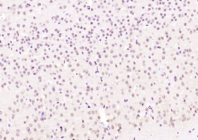
[IF=3.37] Jing Liet al. hUSLCMSCs ameliorated CUMS-induced depression by modulating complement C3 signaling-mediated microglial polarization during astrocyte-microglia crosstalk. Brain Res Bull
. 2020 Oct;163:109-119. IHC ; mouse.
[IF=3.041] An G et al. Effects of CCL5 on the biological behavior of breast cancer and the mechanisms of its interaction with tumor‑associated macrophages. Oncol Rep. 2019 Dec;42(6):2499-2511. IHSLCP ; Human.
[IF=3.943] Yihan Lu et al. Experimental evidence for alpha enolase as one potential autoantigen in the pathogenesis of both autoimmune thyroiditis and its related encephalopathy. Int Immunopharmacol. 2020 Aug;85:106563. IF ; Mouse.
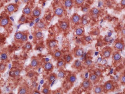
[IF=11.492] Laura Lopez-Sanz. et al. Fcγ receptor activation mediates vascular inflammation and abdominal aortic aneurysm development. Clin Transl Med. 2021 Jul;11(7):e463 IHC ; Human.
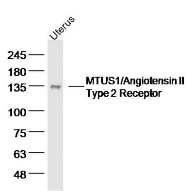
[IF=4.486] Yang Jiao. et al. Inhibition of microglial receptor‐interacting protein kinase 1 ameliorates neuroinflammation following cerebral ischaemic stroke. J Cell Mol Med. 2020 Nov;24(21):12585-12598 WB,IF ; Mouse.
[IF=2.38] Ding, Peng, et al. "Expression of Tumor-Associated Macrophage in Progression of Human Glioma." Cell Biochemistry and Biophysics (2014): 1-7. IHSLCP ; Human.