Home > Product > Antibody > Rabbit Anti-ARHGAP32 antibody
Brain-specific Rho GTPase-activating protein; GAB-associated Cdc42/Rac GTPase-activating protein; GC-GAP; GRIT; GTPase regulator interacting with TrkA; p200RhoGAP; p250GAP; PX-RICS; rac GTPase activating protein; Rho GTPase activating protein 32; Rho GTPa
Cat:
SL9296R
Species Reactivity:
Mouse,Rat,(predicted: Human,Dog,Pig,Cow,Horse,)
Immunogen:
KLH conjugated synthetic peptide derived from human ARHGAP32:801-920/2078
Format:
Liquid
Storage instructions:
Shipped at 4℃. Store at -20 °C for one year. Avoid repeated freeze/thaw cycles.
Concentration:
1mg/ml
Clonality:
Polyclonal
Isotype:
IgG
Applications:
ELISA=1:5000-10000IHC-P=1:100-500IHC-F=1:100-500Flow-Cyt=2ug/TestIF=1:50-200(Paraffin sections need to do antigen repair)not yet tested in other applications.optimal dilutions/concentrations should be determined by the end user.
Host:
Rabbit
Product Overview:
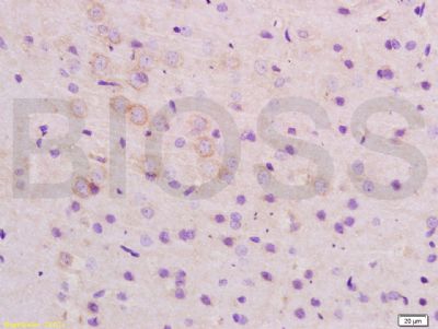
Tissue/cell: rat brain tissue; 4% Paraformaldehyde-fixed and paraffin-embedded; Antigen retrieval: citrate buffer ( 0.01M, pH 6.0 ), Boiling bathing for 15min; Block endogenous peroxidase by 3% Hydrogen peroxide for 30min; Blocking buffer (normal goat serum,SLC0005) at 37℃ for 20 min; Incubation: Anti-ARHGAP32 Polyclonal Antibody, Unconjugated(SL9296R) 1:200, overnight at 4°C, followed by conjugation to the secondary antibody(SP-0023) and DAB(SLC0010) stainingBlank control:Mouse spleen. Primary Antibody (green line): Rabbit Anti-ARHGAP32 antibody (SL9296R) Dilution: 2μg /10^6 cells; Isotype Control Antibody (orange line): Rabbit IgG . Secondary Antibody : Goat anti-rabbit IgG-AF488Dilution: 1μg /test. ProtocolThe cells were fixed with 70% ethanol (10min at room temperature) and then were incubated in 5%BSA to block non-specific protein-protein interactions for 30 min at room temperature .Cells stained with Primary Antibody for 30 min at room temperature. The secondary antibody used for 40 min at room temperature. Acquisition of 20,000 events was performed.
More
Unit:
Price: $
Product PDFs
Datasheet:


ARHGAP32 is a neuron-associated GTPase-activating protein that may regulate dendritic spine morphology and strength by modulating Rho GTPase.

Function:
GTPase-activating protein (GAP) promoting GTP hydrolysis on RHOA, CDC42 and RAC1 small GTPases. May be involved in the differentiation of neuronal cells during the formation of neurite extensions. Involved in NMDA receptor activity-dependent actin reorganization in dendritic spines. May mediate cross-talks between Ras- and Rho-regulated signaling pathways in cell growth regulation. Isoform 2 has higher GAP activity (By similarity).

Subunit:
nteracts with NTRK1 (via cytoplasmic domain); the interaction is independent of the phosphorylation state of NTRK1. Interacts with SHC3 (via SH2 domain). Interacts with RASA1 (via SH3 domain); the interaction is necessary for the Ras activation and cell transforming activities of ARHGAP32 (By similarity). Interacts with GAB1 and GAB2. Interacts with CRK and CRKL. Found in a complex with CRKL and BCAR1; upon EGF stimulation BCAR1 may be replaced by EGFR. Interacts with NCK1 (via SH3 domain); NCK1 recruits phosphorylated BCAR1 to the complex. Isoform 2 interacts with FYN; the interaction appears to be dependent on tyrosine phosphorylation of ARHGAP32. Interacts with EGFR; the interaction requires EGF stimulation and is increased by SHC3. Interacts with CDC42; the interaction requires constitutively active CDC42. Interacts with CTNNB1, DLG4, CDH2 and GRIN2B (By similarity).

Subcellular Location:
Cell junction, synapse, postsynaptic cell membrane, postsynaptic density. Cell projection, dendritic spine By similarity. Cytoplasm, cell cortex. Endosome membrane By similarity. Golgi apparatus membrane By similarity. Endoplasmic reticulum membrane By similarity. Membrane.

Tissue Specificity:
Isoform 1 and isoform 2 are highly expressed in brain and testis. Isoform 1 is also expressed in other tissues such as lung, liver and spleen.

Post-translational modifications:
Isoform 2 is phosphorylated on multiple tyrosine residues by FYN. Phosphorylated tyrosine residues undergo dephosphorylation after stimulation of NMDA receptors (By similarity). Phosphorylated in vitro by CaMK2 in the presence of calmodulin and calcium; which inhibits GAP activity (By similarity).

Similarity:
Belongs to the PX domain-containing GAP family.
Contains 1 PX (phox homology) domain.
Contains 1 Rho-GAP domain.
Contains 1 SH3 domain.

SWISS:
A7KAX9

Gene ID:
9743

Database links:

Entrez Gene: 9743 Human

Omim: 608541 Human

SwissProt: A7KAX9 Human

Unigene: 88379 Human


Picture

Tissue/cell: rat brain tissue; 4% Paraformaldehyde-fixed and paraffin-embedded;
Antigen retrieval: citrate buffer ( 0.01M, pH 6.0 ), Boiling bathing for 15min; Block endogenous peroxidase by 3% Hydrogen peroxide for 30min; Blocking buffer (normal goat serum,SLC0005) at 37℃ for 20 min;
Incubation: Anti-ARHGAP32 Polyclonal Antibody, Unconjugated(SL9296R) 1:200, overnight at 4°C, followed by conjugation to the secondary antibody(SP-0023) and DAB(SLC0010) staining
Blank control:Mouse spleen.
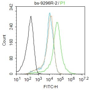
Primary Antibody (green line): Rabbit Anti-ARHGAP32 antibody (SL9296R)
Dilution: 2μg /10^6 cells;
Isotype Control Antibody (orange line): Rabbit IgG .
Secondary Antibody : Goat anti-rabbit IgG-AF488
Dilution: 1μg /test.
Protocol
The cells were fixed with 70% ethanol (10min at room temperature) and then were incubated in 5%BSA to block non-specific protein-protein interactions for 30 min at room temperature .Cells stained with Primary Antibody for 30 min at room temperature. The secondary antibody used for 40 min at room temperature. Acquisition of 20,000 events was performed.
Product Feedback Wall
Message :
Your Email :
Copyright © 2007-2018 Sunlong Medical All Rights Reserved.