Home > Product > Antibody > Rabbit Anti-phospho-LEF1 (Ser42) antibody
LEF1 (phospho S42); LEF1 (phospho Ser42); p-LEF1(phospho S42); p-LEF1(Ser42); lymphoid enhancing factor-1; Transcription factor T cell specific 1 alpha ; DKFZp586H0919; LEF 1; LEF1; Lymphoid enhancer binding factor 1; T cell specific transcription factor
Cat:
SL9842R
Species Reactivity:
Human,Mouse,(predicted: Rat,Chicken,Dog,Pig,Cow,Rabbit,)
Immunogen:
KLH conjugated synthesised phosphopeptide derived from human LEF1 around the phosphorylation site of Ser42:EI(p-S)HP
Format:
Liquid
Storage instructions:
Shipped at 4℃. Store at -20 °C for one year. Avoid repeated freeze/thaw cycles.
Concentration:
1mg/ml
Clonality:
Polyclonal
Isotype:
IgG
Applications:
WB=1:500-2000IHC-P=1:100-500IHC-F=1:100-500IF=1:50-200(Paraffin sections need to do antigen repair)not yet tested in other applications.optimal dilutions/concentrations should be determined by the end user.
Host:
Rabbit
Product Overview:
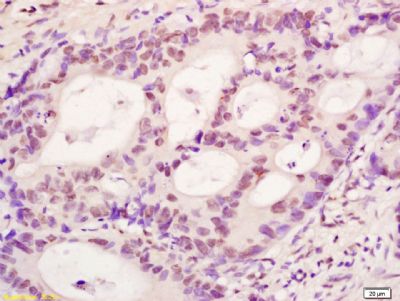
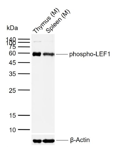
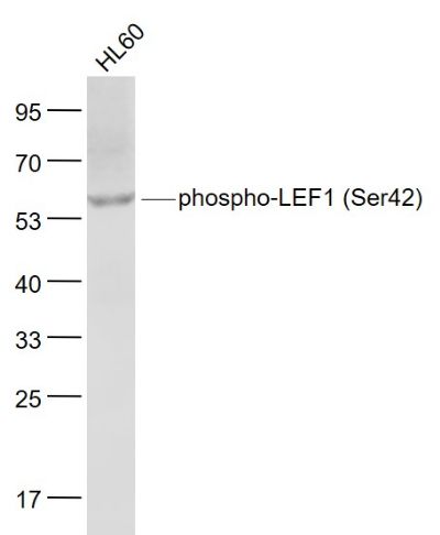
Sample: Lane 1: Mouse Thymus tissue lysates Lane 2: Mouse Spleen tissue lysates Primary: Anti-phospho-LEF1 (Ser42) (SL9842R) at 1/1000 dilutionSecondary: IRDye800CW Goat Anti-Rabbit IgG at 1/20000 dilutionPredicted band size: 44 kDaObserved band size: 58 kDaSample: HL60(Human) Cell Lysate at 30 ugPrimary: Anti- phospho-LEF1 (Ser42) (SL9842R) at 1/1000 dilutionSecondary: IRDye800CW Goat Anti-Rabbit IgG at 1/20000 dilutionPredicted band size: 44 kDObserved band size: 58 kDTissue/cell: human colon carcinoma; 4% Paraformaldehyde-fixed and paraffin-embedded; Antigen retrieval: citrate buffer ( 0.01M, pH 6.0 ), Boiling bathing for 15min; Block endogenous peroxidase by 3% Hydrogen peroxide for 30min; Blocking buffer (normal goat serum,SLC0005) at 37℃ for 20 min; Incubation: Anti-phospho-LEF1(Ser42) Polyclonal Antibody, Unconjugated(SL9842R) 1:200, overnight at 4°C, followed by conjugation to the secondary antibody(SP-0023) and DAB(SLC0010) staining
More
Unit:
Price: $
Product PDFs
Datasheet:


This gene encodes a transcription factor belonging to a family of proteins that share homology with the high mobility group protein-1. The protein encoded by this gene can bind to a functionally important site in the T-cell receptor-alpha enhancer, thereby conferring maximal enhancer activity. This transcription factor is involved in the Wnt signaling pathway, and it may function in hair cell differentiation and follicle morphogenesis. Mutations in this gene have been found in somatic sebaceous tumors. This gene has also been linked to other cancers, including androgen-independent prostate cancer. Alternative splicing results in multiple transcript variants. [provided by RefSeq, Oct 2009].

Function:
Participates in the Wnt signaling pathway. Activates transcription of target genes in the presence of CTNNB1 and EP300. May play a role in hair cell differentiation and follicle morphogenesis. TLE1, TLE2, TLE3 and TLE4 repress transactivation mediated by LEF1 and CTNNB1. Regulates T-cell receptor alpha enhancer function. Binds DNA in a sequence-specific manner. PIAG antagonizes both Wnt-dependent and Wnt-independent activation by LEF1 (By similarity). Isoform 3 lacks the CTNNB1 interaction domain and may be an antagonist for Wnt signaling. Isoform 5 transcriptionally activates the fibronectin promoter, binds to and represses transcription from the E-cadherin promoter in a CTNNB1-independent manner, and is involved in reducing cellular aggregation and increasing cell migration of pancreatic cancer cells. Isoform 1 transcriptionally activates MYC and CCND1 expression and enhances proliferation of pancreatic tumor cells.

Subunit:
Binds the armadillo repeat of CTNNB1 and forms a stable complex. Interacts with EP300, TLE1 and PIASG (By similarity). Binds ALYREF/THOC4, MDFI and MDFIC. Interacts with NLK.

Subcellular Location:
Nucleus. Note=Found in nuclear bodies upon PIASG binding.

Tissue Specificity:
Detected in thymus. Not detected in normal colon, but highly expressed in colon cancer biopsies and colon cancer cell lines. Expressed in several pancreatic tumors and weakly expressed in normal pancreatic tissue. Isoforms 1 and 5 are detected in several pancreatic cell lines.

Post-translational modifications:
Phosphorylated at Thr-155 and/or Ser-166 by NLK. Phosphorylation by NLK at these sites represses LEF1-mediated transcriptional activation of target genes of the canonical Wnt signaling pathway.

Similarity:
Belongs to the TCF/LEF family.
Contains 1 HMG box DNA-binding domain.

SWISS:
Q9UJU2

Gene ID:
51176

Database links:

Entrez Gene: 51176 Human

Omim: 153245 Human

SwissProt: Q9UJU2 Human

Unigene: 726506 Human


lef-1是淋巴增强因子/T-细胞因子(LEF/TCF)家族的成员之一,属于高迁移率组分蛋白家族。lef-1也是一个重要的转录因子,是Wnt信号通路的核心成分。 Picture

Sample:
Lane 1: Mouse Thymus tissue lysates
Lane 2: Mouse Spleen tissue lysates
Primary: Anti-phospho-LEF1 (Ser42) (SL9842R) at 1/1000 dilution
Secondary: IRDye800CW Goat Anti-Rabbit IgG at 1/20000 dilution
Predicted band size: 44 kDa
Observed band size: 58 kDa
Sample:
HL60(Human) Cell Lysate at 30 ug
Primary: Anti- phospho-LEF1 (Ser42) (SL9842R) at 1/1000 dilution
Secondary: IRDye800CW Goat Anti-Rabbit IgG at 1/20000 dilution
Predicted band size: 44 kD
Observed band size: 58 kD
Tissue/cell: human colon carcinoma; 4% Paraformaldehyde-fixed and paraffin-embedded;
Antigen retrieval: citrate buffer ( 0.01M, pH 6.0 ), Boiling bathing for 15min; Block endogenous peroxidase by 3% Hydrogen peroxide for 30min; Blocking buffer (normal goat serum,SLC0005) at 37℃ for 20 min;
Incubation: Anti-phospho-LEF1(Ser42) Polyclonal Antibody, Unconjugated(SL9842R) 1:200, overnight at 4°C, followed by conjugation to the secondary antibody(SP-0023) and DAB(SLC0010) staining
Product Feedback Wall
Message :
Your Email :
Copyright © 2007-2018 Sunlong Medical All Rights Reserved.