OLFM3 is a 478 amino acid protein that interacts with myocilin. Myocilin is an extracellular protein that plays a key role in the actomyosin system and is responsible for controlling intraocular pressure. OLFM3 is a secreted protein that contains an olfactomedin-like (OLF) domain, an approximately 260 amino acid motif commonly found in secreted glycoproteins. OLFM3 localizes to the Golgi apparatus of the cell and is highly expressed in both eye and brain tissue. Mutations in the gene that encodes OLFM3 may cause severe glaucoma, a condition in which increased intraocular pressure within the eyeball causes loss of eye sight.
Subunit:
Peripherally associated with AMPAR complex. AMPAR complex consists of an inner core made of 4 pore-forming GluA/GRIA proteins (GRIA1, GRIA2, GRIA3 and GRIA4) and 4 major auxiliary subunits arranged in a twofold symmetry. One of the two pairs of distinct binding sites is occupied either by CNIH2, CNIH3 or CACNG2, CACNG3. The other harbors CACNG2, CACNG3, CACNG4, CACNG8 or GSG1L. This inner core of AMPAR complex is complemented by outer core constituents binding directly to the GluA/GRIA proteins at sites distinct from the interaction sites of the inner core constituents. Outer core constituents include at least PRRT1, PRRT2, CKAMP44/SHISA9, FRRS1L and NRN1. The proteins of the inner and outer core serve as a platform for other, more peripherally associated AMPAR constituents, including OLFM3. Alone or in combination, these auxiliary subunits control the gating and pharmacology of the AMPAR complex and profoundly impact their biogenesis and protein processing. Homodimer. Interacts with MYOC (By similarity).
Subcellular Location:
Secreted.
Tissue Specificity:
In the eye, expressed in trabecular meshwork and neural retina; in non-ocular tissues, expressed in brain and lung.
Similarity:
Contains 1 olfactomedin-like domain.
SWISS:
Q96PB7
Gene ID:
118427
Database links:
Entrez Gene: 118427 Human
Entrez Gene: 229759 Mouse
Entrez Gene: 252920 Rat
Omim: 607567 Human
SwissProt: Q96PB7 Human
SwissProt: P63056 Mouse
SwissProt: P63057 Rat
Unigene: 484475 Human
Unigene: 54183 Mouse
Unigene: 27711 Rat
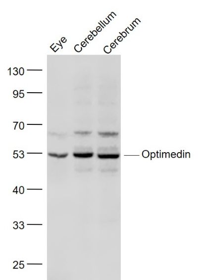
| Picture |
Sample:
Eye (Mouse) Lysate at 40 ug
Cerebellum (Mouse) Lysate at 40 ug
Cerebrum (Mouse) Lysate at 40 ug
Primary: Anti- Optimedin (SL11061R) at 1/1000 dilution
Secondary: IRDye800CW Goat Anti-Rabbit IgG at 1/20000 dilution
Predicted band size: 52 kD
Observed band size: 52 kD
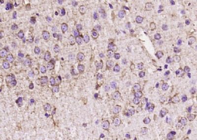
Paraformaldehyde-fixed, paraffin embedded (rat brain); Antigen retrieval by boiling in sodium citrate buffer (pH6.0) for 15min; Block endogenous peroxidase by 3% hydrogen peroxide for 20 minutes; Blocking buffer (normal goat serum) at 37°C for 30min; Antibody incubation with (Optimedin) Polyclonal Antibody, Unconjugated (SL11061R) at 1:200 overnight at 4°C, followed by operating according to SP Kit(Rabbit) (sp-0023) instructionsand DAB staining.
|
|
|