Home > Product > Antibody > Rabbit Anti-Complement component C9a antibody
C9 deficiency; C9a; C9 deficiency with dermatomyositis; CO9_HUMAN; Complement component 9; Complement component 9 deficiency; Complement component C9a; Complement component C9a; C9.
Cat:
SL11190R
Species Reactivity:
Human,
Immunogen:
KLH conjugated synthetic peptide derived from human Complement component C9a:161-260/559
Format:
Liquid
Storage instructions:
Shipped at 4℃. Store at -20 °C for one year. Avoid repeated freeze/thaw cycles.
Concentration:
1mg/ml
Clonality:
Polyclonal
Isotype:
IgG
Applications:
WB=1:500-2000ELISA=1:5000-10000Flow-Cyt=3ug/Testnot yet tested in other applications.optimal dilutions/concentrations should be determined by the end user.
Host:
Rabbit
Product Overview:
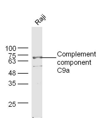
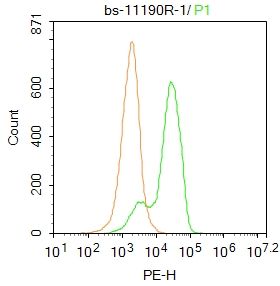
Sample: Raji Cell (Human) Lysate at 30 ugPrimary: Anti- Complement component C9a (Bs- 11190R) at 1/300 dilutionSecondary: IRDye800CW Goat Anti-Rabbit IgG at 1/20000 dilutionPredicted band size: 26/61 kDObserved band size: 70 kDBlank control: HL60. Primary Antibody (green line): Rabbit Anti-Complement component C9a antibody (SL11190R) Dilution: 1μg /10^6 cells; Isotype Control Antibody (orange line): Rabbit IgG . Secondary Antibody : Goat anti-rabbit IgG-PEDilution: 1μg /test. ProtocolThe cells then incubated in 5%BSA to block non-specific protein-protein interactions for 30 min at at room temperature .Cells stained with Primary Antibody for 30 min at room temperature. The secondary antibody used for 40 min at room temperature. Acquisition of 20,000 events was performed.
More
Unit:
Price: $
Product PDFs
Datasheet:


C9 is a plasma protein synthesized in the liver and monocytes consisting of a single polypeptide chain. C9 is a part of the membrane attack complex (MAC), an important component of the immune system. The MAC forms upon complement system activation by invading pathogenic bacteria and consists of the four major complement proteins: C5b, C6, C7 and C8. These complement proteins bind to the outer surface of the plasma membrane of the invading cell. C9 binds to the membrane associated C5b-8 protein, which leads to the circular polymerization of 12-18 C9 molecules. These polymerized C9 molecules form a ring structure in the membrane. Molecules can then diffuse freely through this transmembrane channel, causing cell lysis and destruction of the invading bacterial cell.

Function:
Constituent of the membrane attack complex (MAC) that plays a key role in the innate and adaptive immune response by forming pores in the plasma membrane of target cells. C9 is the pore-forming subunit of the MAC.

Subunit:
Component of the membrane attack complex (MAC). MAC assembly is initiated by protelytic cleavage of C5 into C5a and C5b. C5b binds sequentially C6, C7, C8 and multiple copies of the pore-forming subunit C9.

Subcellular Location:
Secreted. Cell membrane; Multi-pass membrane protein. Note=Secreted as soluble monomer. Oligomerizes at target membranes, forming a pre-pore. A conformation change then leads to the formation of a 100 Angstrom diameter pore.

Tissue Specificity:
Plasma.

Post-translational modifications:
Thrombin cleaves factor C9 to produce C9a and C9b.
Phosphorylation sites are present in the extracellular medium.

DISEASE:
Defects in C9 are a cause of complement component 9 deficiency (C9D) [MIM:613825]. A rare defect of the complement classical pathway associated with susceptibility to severe recurrent infections, predominantly by Neisseria gonorrhoeae or Neisseria meningitidis.

Similarity:
Belongs to the complement C6/C7/C8/C9 family.
Contains 1 EGF-like domain.
Contains 1 LDL-receptor class A domain.
Contains 1 MACPF domain.
Contains 1 TSP type-1 domain.

SWISS:
P02748

Gene ID:
735

Database links:

Entrez Gene: 735 Human

Omim: 120940 Human

SwissProt: P02748 Human

Unigene: 654443 Human


Picture

Sample:
Raji Cell (Human) Lysate at 30 ug
Primary: Anti- Complement component C9a (Bs- 11190R) at 1/300 dilution
Secondary: IRDye800CW Goat Anti-Rabbit IgG at 1/20000 dilution
Predicted band size: 26/61 kD
Observed band size: 70 kD
Blank control: HL60.
Primary Antibody (green line): Rabbit Anti-Complement component C9a antibody (SL11190R)
Dilution: 1μg /10^6 cells;
Isotype Control Antibody (orange line): Rabbit IgG .
Secondary Antibody : Goat anti-rabbit IgG-PE
Dilution: 1μg /test.
Protocol
The cells then incubated in 5%BSA to block non-specific protein-protein interactions for 30 min at at room temperature .Cells stained with Primary Antibody for 30 min at room temperature. The secondary antibody used for 40 min at room temperature. Acquisition of 20,000 events was performed.
Product Feedback Wall
Message :
Your Email :
Copyright © 2007-2018 Sunlong Medical All Rights Reserved.