Home > Product > Antibody > Rabbit Anti-ABCG2 antibody
ABC transporter; ABC15; ABCG 2; ABCG2; ABCG2_HUMAN; ABCP; ATP binding cassette sub family G (WHITE) member 2; ATP binding cassette transporter G2; ATP-binding cassette sub-family G member 2; BCRP; BCRP1; BMDP; Breast cancer resistance protein;
Cat:
SL4780R
Species Reactivity:
Human,Rat,(predicted: Mouse,Dog,Horse,)
Immunogen:
KLH conjugated synthetic peptide derived from human ABCG2:561-655/655<Extracellular>
Format:
Liquid
Storage instructions:
Shipped at 4℃. Store at -20 °C for one year. Avoid repeated freeze/thaw cycles.
Concentration:
1mg/ml
Clonality:
Polyclonal
Isotype:
IgG
Applications:
ELISA=1:5000-10000IHC-P=1:100-500IHC-F=1:100-500Flow-Cyt=3ug/testICC=1:100IF=1:100-500(Paraffin sections need to do antigen repair)not yet tested in other applications.optimal dilutions/concentrations should be determined by the end user.
Host:
Rabbit
Product Overview:
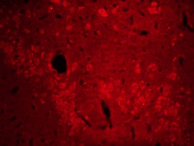
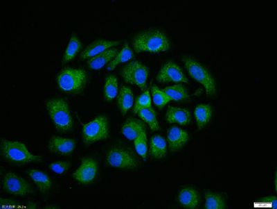
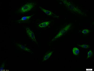
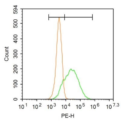
Tissue/cell: rat brain tissue;4% Paraformaldehyde-fixed and paraffin-embedded; Antigen retrieval: citrate buffer ( 0.01M, pH 6.0 ), Boiling bathing for 15min; Blocking buffer (normal goat serum,SLC0005) at 37℃ for 20 min; Incubation: Anti-ABCG2 Polyclonal Antibody, Unconjugated(SL4780R) 1:200, overnight at 4°C; The secondary antibody was Goat Anti-Rabbit IgG, Cy3 conjugated(SL0295G-Cy3)used at 1:200 dilution for 40 minutes at 37°C. HepG2 cell; 4% Paraformaldehyde-fixed; Triton X-100 at room temperature for 20 min; Blocking buffer (normal goat serum, SLC0005) at 37°C for 20 min; Antibody incubation with (ABCG2) polyclonal Antibody, Unconjugated (SL4780R) 1:100, 90 minutes at 37°C; followed by a conjugated Goat Anti-Rabbit IgG antibody at 37°C for 90 minutes, DAPI (blue, C02-04002) was used to stain the cell nuclei.Tissue/cell:A549 cell; 4% Paraformaldehyde-fixed; Triton X-100 at room temperature for 20 min; Blocking buffer (normal goat serum,SLC0005) at 37°C for 20 min; Antibody incubation with (ABCG2) polyclonal Antibody, Unconjugated (SL4780R) 1:100, 90 minutes at 37°C; followed by a FITC conjugated Goat Anti-Rabbit IgG antibody at 37°C for 90 minutes, DAPI (blue, C02-04002) was used to stain the cell nuclei.Blank control: A549. Primary Antibody (green line): Rabbit Anti-ABCG2 antibody (SL4780R) Dilution: 3μg /10^6 cells; Isotype Control Antibody (orange line): Rabbit IgG . Secondary Antibody : Goat anti-rabbit IgG-PEDilution: 3μg /test. ProtocolThe cells were incubated in 5%BSA to block non-specific protein-protein interactions for 30 min at at room temperature .Cells stained with Primary Antibody for 30 min at room temperature. The secondary antibody used for 40 min at room temperature. Acquisition of 20,000 events was performed.
More
Unit:
Price: $
Product PDFs
Datasheet:


An ABC transporter. Allows efflux of Hoechst dye, a property that has been used to separate bone marrow side population cells, which express BCRP/ABCG2. Appears to play a major role in the multidrug resistance phenotype of a specific MCF-7 breast cancer cell line. When overexpressed, the transfected cells become resistant to mitoxantrone, daunorubicin and doxorubicin, display diminished intracellular accumulation of daunorubicin, and manifest an ATP-dependent increase in the efflux of rhodamine 123.
Breast Cancer Resistance Protein (BCRP) is a 70 kDa ATP-Binding Cassette membrane transport protein involved in multidrug resistance. BCRP may be over-expressed in cancer cell lines selected with doxorubicin / verapamil, topotecan or mitoxantrone.


Function:
Xenobiotic transporter that may play an important role in the exclusion of xenobiotics from the brain. May be involved in brain-to-blood efflux. Appears to play a major role in the multidrug resistance phenotype of several cancer cell lines. When overexpressed, the transfected cells become resistant to mitoxantrone, daunorubicin and doxorubicin, display diminished intracellular accumulation of daunorubicin, and manifest an ATP-dependent increase in the efflux of rhodamine 123.

Subunit:
Monomer or homodimer; disulfide-linked.

Subcellular Location:
Cell membrane; Multi-pass membrane protein.

Tissue Specificity:
Highly expressed in placenta. Low expression in small intestine, liver and colon.

Similarity:
Belongs to the ABC transporter superfamily. ABCG family. Eye pigment precursor importer (TC 3.A.1.204) subfamily.
Contains 1 ABC transmembrane type-2 domain.
Contains 1 ABC transporter domain.

SWISS:
Q9UNQ0

Gene ID:
9429

Database links:

Entrez Gene: 9429 Human

Entrez Gene: 26357 Mouse

Entrez Gene: 397073 Pig

Entrez Gene: 312382 Rat

Omim: 603756 Human

SwissProt: Q9UNQ0 Human

SwissProt: Q7TMS5 Mouse

SwissProt: Q8MIB3 Pig

SwissProt: Q80W57 Rat

Unigene: 96218 Human

Unigene: 333096 Mouse

Unigene: 13131 Rat


Picture

Tissue/cell: rat brain tissue;4% Paraformaldehyde-fixed and paraffin-embedded;
Antigen retrieval: citrate buffer ( 0.01M, pH 6.0 ), Boiling bathing for 15min; Blocking buffer (normal goat serum,SLC0005) at 37℃ for 20 min;
Incubation: Anti-ABCG2 Polyclonal Antibody, Unconjugated(SL4780R) 1:200, overnight at 4°C; The secondary antibody was Goat Anti-Rabbit IgG, Cy3 conjugated(SL0295G-Cy3)used at 1:200 dilution for 40 minutes at 37°C.
HepG2 cell; 4% Paraformaldehyde-fixed; Triton X-100 at room temperature for 20 min; Blocking buffer (normal goat serum, SLC0005) at 37°C for 20 min; Antibody incubation with (ABCG2) polyclonal Antibody, Unconjugated (SL4780R) 1:100, 90 minutes at 37°C; followed by a conjugated Goat Anti-Rabbit IgG antibody at 37°C for 90 minutes, DAPI (blue, C02-04002) was used to stain the cell nuclei.
Tissue/cell:A549 cell; 4% Paraformaldehyde-fixed; Triton X-100 at room temperature for 20 min; Blocking buffer (normal goat serum,SLC0005) at 37°C for 20 min; Antibody incubation with (ABCG2) polyclonal Antibody, Unconjugated (SL4780R) 1:100, 90 minutes at 37°C; followed by a FITC conjugated Goat Anti-Rabbit IgG antibody at 37°C for 90 minutes, DAPI (blue, C02-04002) was used to stain the cell nuclei.
Blank control: A549.
Primary Antibody (green line): Rabbit Anti-ABCG2 antibody (SL4780R)
Dilution: 3μg /10^6 cells;
Isotype Control Antibody (orange line): Rabbit IgG .
Secondary Antibody : Goat anti-rabbit IgG-PE
Dilution: 3μg /test.
Protocol
The cells were incubated in 5%BSA to block non-specific protein-protein interactions for 30 min at at room temperature .Cells stained with Primary Antibody for 30 min at room temperature. The secondary antibody used for 40 min at room temperature. Acquisition of 20,000 events was performed.
Product Feedback Wall
Message :
Your Email :
Copyright © 2007-2018 Sunlong Medical All Rights Reserved.