The photoreceptor rod cell that is responsible for vision under conditions of low light consists of stacked arrays of disk membranes that make up its outer segment portion. Regulated by complex biochemical mechanisms, the rod outer segment is under constant renewal as new disks form at the base. MREG (melanoregulin), also known as DSU (dilute suppressor protein homolog) or WDT2, is thought to play a role in membrane fusion and in regulating the biogenesis of disk membranes of photoreceptor rods. MREG interacts with RDS (also known as Peripherin-2), a photoreceptor specific tetraspanin protein that is required to maintain normal cell structure during the renewal process of membrane fusion. MREG is 214 amino acids in length, is expressed in photoreceptor cells and and is expressed as two isoforms due to alternative splicing.
Function:
DSU (Dilute suppressor) is also known as Melanoregulin. It has a role in the incorporation of pigments into hair. It is involved in organelle biogenesis and may function in membrane fusion. It may also regulate the biogenesis of disk membranes, specialized organelles of photoreceptor rod cells.
Subcellular Location:
Apical cell membrane; Peripheral membrane protein. Note: Localizes to the inner segment and basal outer segment of rods in the retina.
Tissue Specificity:
Expressed in photoreceptor cells (at protein level).
Similarity:
Belongs to the melanoregulin family.
SWISS:
Q8N565
Gene ID:
55686
Database links:
Entrez Gene: 55686 Human
Entrez Gene: 381269 Mouse
Omim: 609207 Human
SwissProt: Q8N565 Human
SwissProt: Q6NVG5 Mouse
| Picture |
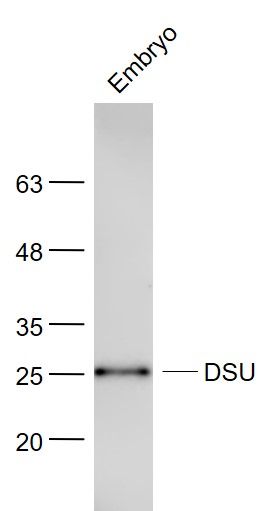
Sample:
Embryo (Mouse) Lysate at 40 ug
Primary: Anti- DSU (SL14433R) at 1/300 dilution
Secondary: IRDye800CW Goat Anti-Rabbit IgG at 1/20000 dilution
Predicted band size: 25 kD
Observed band size: 25 kD
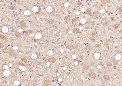
Paraformaldehyde-fixed, paraffin embedded (rat brain); Antigen retrieval by boiling in sodium citrate buffer (pH6.0) for 15min; Block endogenous peroxidase by 3% hydrogen peroxide for 20 minutes; Blocking buffer (normal goat serum) at 37°C for 30min; Antibody incubation with (DSU) Polyclonal Antibody, Unconjugated (SL14433R) at 1:200 overnight at 4°C, followed by operating according to SP Kit(Rabbit) (sp-0023) instructionsand DAB staining.
|
|
|