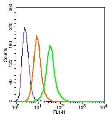
Blank control:H9C2 Cells(blue).
Primary Antibody: Rabbit Anti-CANX/FITC Conjugated antibody (SL1693R/FITC), Dilution: 1μg in 100 μL 1X PBS containing 0.5% BSA;
Isotype Control Antibody: Rabbit IgG/FITC(orange) ,used under the same conditions.
Protocol
The cells were washed twice with phosphate-buffered saline (PBS). The cells were incubated in 1 X PBS containing 0.5% BSA + 1 0% goat serum (15 min) to block non-specific protein-protein interactions followed by the antibody (SL1693R/FITC, 1μg /1x10^6 cells) for 30 min on ice. Acquisition of 20,000 events was performed.