Home > Product > Antibody > Rabbit Anti-EPS8 antibody
Epidermal growth factor receptor kinase substrate 8; Epidermal growth factor receptor pathway substrate 8; EPS 8; EPS-8; EPS8_HUMAN.
Cat:
SL3848R
Species Reactivity:
Human,Mouse,(predicted: Rat,Dog,Pig,Cow,Horse,Guinea Pig,)
Immunogen:
KLH conjugated synthetic peptide derived from human EPS8:251-350/822
Format:
Liquid
Storage instructions:
Shipped at 4℃. Store at -20 °C for one year. Avoid repeated freeze/thaw cycles.
Concentration:
1mg/ml
Clonality:
Polyclonal
Isotype:
IgG
Applications:
WB=1:500-2000ELISA=1:5000-10000IHC-P=1:100-500IHC-F=1:100-500IF=1:100-500(Paraffin sections need to do antigen repair)not yet tested in other applications.optimal dilutions/concentrations should be determined by the end user.
Host:
Rabbit
Product Overview:
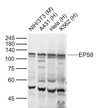
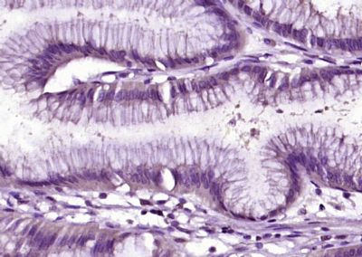
Sample: Lane 1: NIH/3T3 (Mouse) Cell Lysate at 30 ugLane 2: A431 (Human) Cell Lysate at 30 ugLane 3: Hela (Human) Cell Lysate at 30 ugLane 4: K562 (Human) Cell Lysate at 30 ugPrimary: Anti-EPS8 (SL3848R) at 1/1000 dilutionSecondary: IRDye800CW Goat Anti-Rabbit IgG at 1/20000 dilutionPredicted band size: 92 kDObserved band size: 120 kDParaformaldehyde-fixed, paraffin embedded (human gastric carcinoma); Antigen retrieval by boiling in sodium citrate buffer (pH6.0) for 15min; Block endogenous peroxidase by 3% hydrogen peroxide for 20 minutes; Blocking buffer (normal goat serum) at 37°C for 30min; Antibody incubation with (EPS8) Polyclonal Antibody, Unconjugated (SL3848R) at 1:200 overnight at 4°C, followed by operating according to SP Kit(Rabbit) (sp-0023) instructionsand DAB staining.
More
Unit:
Price: $
Product PDFs
Datasheet:


This gene encodes a member of the EPS8 family. This protein contains one PH domain and one SH3 domain. It functions as part of the EGFR pathway, though its exact role has not been determined. Highly similar proteins in other organisms are involved in the transduction of signals from Ras to Rac and growth factor-mediated actin remodeling. Alternate transcriptional splice variants of this gene have been observed but have not been thoroughly characterized. [provided by RefSeq, Jul 2008].

Function:
Signaling adapter that controls various cellular protrusions by regulating actin cytoskeleton dynamics and architecture. Depending on its association with other signal transducers, can regulate different processes. Together with SOS1 and ABI1, forms a trimeric complex that participates in transduction of signals from Ras to Rac by activating the Rac-specific guanine nucleotide exchange factor (GEF) activity. Acts as a direct regulator of actin dynamics by binding actin filaments and has both barbed-end actin filament capping and actin bundling activities depending on the context. Displays barbed-end actin capping activity when associated with ABI1, thereby regulating actin-based motility process: capping activity is auto-inhibited and inhibition is relieved upon ABI1 interaction. Also shows actin bundling activity when associated with BAIAP2, enhancing BAIAP2-dependent membrane extensions and promoting filopodial protrusions. Involved in the regulation of processes such as axonal filopodia growth, stereocilia length, dendritic cell migration and cancer cell migration and invasion. Acts as a regulator of axonal filopodia formation in neurons: in the absence of neurotrophic factors, negatively regulates axonal filopodia formation via actin-capping activity. In contrast, it is phosphorylated in the presence of BDNF leading to inhibition of its actin-capping activity and stimulation of filopodia formation. Component of a complex with DFNB31 and MYO15A that localizes at stereocilia tips and is required for elongation of the stereocilia actin core. Indirectly involved in cell cycle progression; its degradation following ubiquitination being required during G2 phase to promote cell shape changes.

Subunit:
Homodimer. Part of a complex consisting of ABI1, EPS8 and SOS1. Interacts with MYO15A and DFNB31. Interacts with LANCL1. Interacts with EGFR; mediates EPS8 phosphorylation. Interacts with BAIAP2. Interacts with SHB.

Subcellular Location:
Cytoplasm, cell cortex. Cell projection, ruffle membrane. Cell projection, growth cone. Cell projection, stereocilium. Cell junction, synapse, synaptosome. Note=Localizes at the tips of the stereocilia of the inner and outer hair cells. Localizes to the midzone of dividing cells.

Tissue Specificity:
Expressed in all tissues analyzed, including heart, brain, placenta, lung, liver, skeletal muscle, kidney and pancreas. Expressed in all epithelial and fibroblastic lines examined and in some, but not all, hematopoietic cells.

Post-translational modifications:
Ubiquitinated by the SCF(FBXW5) E3 ubiquitin-protein ligase complex during G2 phase, leading to its transient degradation and subsequent cell shape changes required to allow mitotic progression. Reappears at the midzone of dividing cells.
Phosphorylation at Ser-625 and Thr-629 by MAPK following BDNF treatment promotes removal from actin and filopodia formation. Phosphorylated by several receptor tyrosine kinases.

DISEASE:
Deafness, autosomal recessive, 102 (DFNB102) [MIM:615974]: A form of non-syndromic deafness characterized by profound hearing loss affecting all frequencies. Vestibular function is unaffected. Note=The disease is caused by mutations affecting the gene represented in this entry.
Note=Defects in EPS8 are associated with some cancers, such as pancreatic, oral squamous cell carcinomas or pituitary cancers. Contributes to cell transformation in response to growth factor treatment and is overexpressed in a number of tumors, indicating that EPS8 levels must be tightly regulated.

Similarity:
Belongs to the EPS8 family.
Contains 1 PH domain.
Contains 1 SH3 domain.

SWISS:
Q12929

Gene ID:
2059

Database links:

Entrez Gene: 2059 Human

Omim: 600206 Human

SwissProt: Q12929 Human

Unigene: 591160 Human


Picture

Sample:
Lane 1: NIH/3T3 (Mouse) Cell Lysate at 30 ug
Lane 2: A431 (Human) Cell Lysate at 30 ug
Lane 3: Hela (Human) Cell Lysate at 30 ug
Lane 4: K562 (Human) Cell Lysate at 30 ug
Primary: Anti-EPS8 (SL3848R) at 1/1000 dilution
Secondary: IRDye800CW Goat Anti-Rabbit IgG at 1/20000 dilution
Predicted band size: 92 kD
Observed band size: 120 kD
Paraformaldehyde-fixed, paraffin embedded (human gastric carcinoma); Antigen retrieval by boiling in sodium citrate buffer (pH6.0) for 15min; Block endogenous peroxidase by 3% hydrogen peroxide for 20 minutes; Blocking buffer (normal goat serum) at 37°C for 30min; Antibody incubation with (EPS8) Polyclonal Antibody, Unconjugated (SL3848R) at 1:200 overnight at 4°C, followed by operating according to SP Kit(Rabbit) (sp-0023) instructionsand DAB staining.
Product Feedback Wall
Message :
Your Email :
Copyright © 2007-2018 Sunlong Medical All Rights Reserved.