Docking protein 1 is constitutively tyrosine phosphorylated in hematopoietic progenitors isolated from chronic myelogenous leukemia (CML) patients in the chronic phase. It may be a critical substrate for p210(bcr/abl), a chimeric protein whose presence is associated with CML. Docking protein 1 contains a putative pleckstrin homology domain at the amino terminus and ten PXXP SH3 recognition motifs. Docking protein 2 binds p120 (RasGAP) from CML cells. It has been postulated to play a role in mitogenic signaling.
Function: DOK proteins are enzymatically inert adaptor or scaffolding proteins. They provide a docking platform for the assembly of multimolecular signaling complexes. DOK1 appears to be a negative regulator of the insulin signaling pathway. Modulates integrin activation by competing with talin for the same binding site on ITGB3.
Subunit: Interacts with ABL1 (By similarity). Interacts with RasGAP and INPP5D/SHIP1. Interacts directly with phosphorylated ITGB3.
Tissue Specificity: Expressed in pancreas, heart, leukocyte and spleen. Expressed in both resting and activated peripheral blood T-cells.
Post-translational modifications: Constitutively tyrosine-phosphorylated. Phosphorylated by TEC. Phosphorylated by LYN. Phosphorylated on tyrosine residues by the insulin receptor kinase. Results in the negative regulation of the insulin signaling pathway. Isoform 3 contains a N-acetylmethionine at position 1.
Similarity: Belongs to the DOK family. Type A subfamily. Contains 1 IRS-type PTB domain. Contains 1 PH domain.
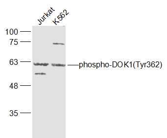
Sample:
Jurkat(Human) Cell Lysate at 30 ug
K562(Human) Cell Lysate at 30 ug
Primary: Anti-phospho-DOK1(Tyr362) (SL5289R) at 1/1000 dilution
Secondary: IRDye800CW Goat Anti-Rabbit IgG at 1/20000 dilution
Predicted band size: 62 kD
Observed band size: 62 kD