DOK1 (phospho Y398); DOK1 (phospho Tyr398); p-DOK1 (phospho Y398); docking protein 1 (downstream of tyrosine kinase 1); Docking protein 1 (p62(dok)) (Downstream of tyrosine kinase 1) (pp62).; docking protein 1 62kD (downstream of tyrosine kinase 1); Docki
Cat:
SL5290R
Species Reactivity:
Mouse,(predicted: Human,Rat,Dog,Pig,Cow,Horse,)
Immunogen:
KLH conjugated Synthesised phosphopeptide derived from human DOK1 around the phosphorylation site of Tyr398:EG(-Y)EL
Format:
Liquid
Storage instructions:
Shipped at 4℃. Store at -20 °C for one year. Avoid repeated freeze/thaw cycles.
Concentration:
1mg/ml
Clonality:
Polyclonal
Isotype:
IgG
Applications:
WB=1:500-2000IHC-P=1:100-500IHC-F=1:100-500IF=1:100-500(Paraffin sections need to do antigen repair)not yet tested in other applications.optimal dilutions/concentrations should be determined by the end user.
Host:
Rabbit
Product Overview:
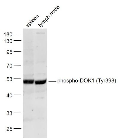
Sample: Spleen (Mouse) Lysate at 40 ugLymph node (Mouse) Lysate at 40 ugPrimary: Anti- phospho-DOK1 (Tyr398) (SL5290R) at 1/1000 dilutionSecondary: IRDye800CW Goat Anti-Rabbit IgG at 1/20000 dilutionPredicted band size: 53 kDObserved band size: 52 kD
Docking protein 1 is constitutively tyrosine phosphorylated in hematopoietic progenitors isolated from chronic myelogenous leukemia (CML) patients in the chronic phase. It may be a critical substrate for p210(bcr/abl), a chimeric protein whose presence is associated with CML. Docking protein 1 contains a putative pleckstrin homology domain at the amino terminus and ten PXXP SH3 recognition motifs. Docking protein 2 binds p120 (RasGAP) from CML cells. It has been postulated to play a role in mitogenic signaling.
Function: DOK proteins are enzymatically inert adaptor or scaffolding proteins. They provide a docking platform for the assembly of multimolecular signaling complexes. DOK1 appears to be a negative regulator of the insulin signaling pathway. Modulates integrin activation by competing with talin for the same binding site on ITGB3.
Subunit: Interacts with ABL1. Interacts with RasGAP and INPP5D/SHIP1. Interacts directly with phosphorylated ITGB3.
Tissue Specificity: Expressed in pancreas, heart, leukocyte and spleen. Expressed in both resting and activated peripheral blood T-cells.
Post-translational modifications: Constitutively tyrosine-phosphorylated. Phosphorylated by TEC (By similarity). Phosphorylated by LYN. Phosphorylated on tyrosine residues by the insulin receptor kinase. Results in the negative regulation of the insulin signaling pathway. Isoform 3 contains a N-acetylmethionine at position 1.
Similarity: Belongs to the DOK family. Type A subfamily. Contains 1 IRS-type PTB domain. Contains 1 PH domain.
Sample:
Spleen (Mouse) Lysate at 40 ug
Lymph node (Mouse) Lysate at 40 ug
Primary: Anti- phospho-DOK1 (Tyr398) (SL5290R) at 1/1000 dilution
Secondary: IRDye800CW Goat Anti-Rabbit IgG at 1/20000 dilution
Predicted band size: 53 kD
Observed band size: 52 kD