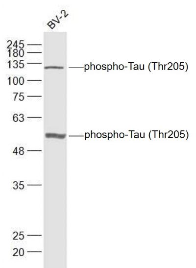
Sample:
BSLV2(Mouse) Cell Lysate at 30 ug
Primary: Anti-phospho-Tau (Thr205) (SL5420R) at 1/1000 dilution
Secondary: IRDye800CW Goat Anti-Rabbit IgG at 1/20000 dilution
Predicted band size: 52/79 kD
Observed band size: 52/129 kD
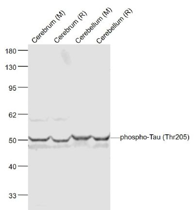
Sample:
Cerebrum (Mouse) Lysate at 40 ug
Cerebrum (Rat) Lysate at 40 ug
Cerebellum (Mouse) Lysate at 40 ug
Cerebellum (Rat) Lysate at 40 ug
Primary: Anti-phospho-Tau (Th205) (SL5420R) at 1/1000 dilution
Secondary: IRDye800CW Goat Anti-Rabbit IgG at 1/20000 dilution
Predicted band size: 50-70 kD
Observed band size: 50 kD
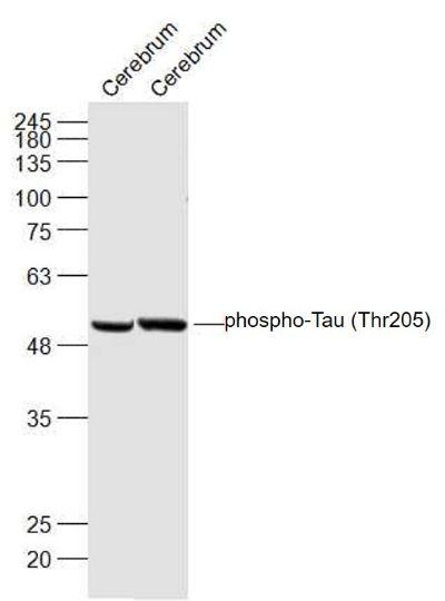
Sample:
Cerebrum (Mouse) Lysate at 40 ug
Cerebrum (Rat) Lysate at 40 ug
Primary: Anti-phospho-Tau (Thr205) (SL5420R) at 1/1000 dilution
Secondary: IRDye800CW Goat Anti-Rabbit IgG at 1/20000 dilution
Predicted band size: 52/79 kD
Observed band size: 52 kD
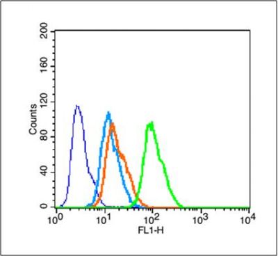
Blank control (blue line): MCF 7 (blue).
Primary Antibody (green line): Rabbit Anti- phospho-Tau (Thr205) antibody (SL5420R)
Dilution: 1μg /10^5 cells;
Isotype Control Antibody (orange line): Rabbit IgG .
Secondary Antibody (white blue line): Goat anti-rabbit IgG-FITC
Dilution: 1μg /test.
Protocol
The cells were fixed with 70% methanol (Overnight at 4℃) and then permeabilized with 90% ice-cold methanol for 30 min on ice. Cells stained with Primary Antibody for 30 min at room temperature. The cells were then incubated in 1 X PBS/2%BSA/10% goat serum to block non-specific protein-protein interactions followed by the antibody for 15 min at room temperature. The secondary antibody used for 40 min at room temperature. Acquisition of 20,000 events was performed.
|