Home > Product > Antibody > Rabbit Anti-phospho-Ron (Ser1349) antibody
MST1R(phospho S1349); c met related tyrosine kinase; CD136; CD136 antigen; CDw136; Macrophage stimulating 1 receptor (c met related tyrosine kinase); Macrophage stimulating 1 receptor; Macrophage stimulating protein receptor alpha chain; MACROPHAGE STIMUL
Cat:
SL5489R
Species Reactivity:
(predicted: Mouse,)
Immunogen:
KLH conjugated Synthesised phosphopeptide derived from mouse MST1R around the phosphorylation site of Ser1349:DG(p-S)VP
Format:
Liquid
Storage instructions:
Shipped at 4℃. Store at -20 °C for one year. Avoid repeated freeze/thaw cycles.
Concentration:
1mg/ml
Clonality:
Polyclonal
Isotype:
IgG
Applications:
WB=1:500-2000ELISA=1:5000-10000IHC-P=1:100-500IHC-F=1:100-500Flow-Cyt=3ug/TestIF=1:100-500(Paraffin sections need to do antigen repair)not yet tested in other applications.optimal dilutions/concentrations should be determined by the end user.
Host:
Rabbit
Product Overview:
Blank control (Black line): A431 (Black). Primary Antibody (green line): Rabbit Anti-phospho-Ron(Ser1349) antibody (SL5489R) Dilution: 3μg /10^6 cells; Isotype Control Antibody (orange line): Rabbit IgG . Secondary Antibody (white blue line): Goat anti-rabbit IgG-AF647Dilution: 3μg /test. ProtocolThe cells were fixed with 4% PFA (10min at room temperature)and then permeabilized with 90% ice-cold methanol for 20 min at room temperature. The cells were then incubated in 5%BSA to block non-specific protein-protein interactions for 30 min at room temperature .Cells stained with Primary Antibody for 30 min at room temperature. The secondary antibody used for 40 min at room temperature. Acquisition of 20,000 events was performed.
More
Unit:
Price: $
Product PDFs
Datasheet:


MST1R/Ron, a HGF Receptor/MET-type protein kinase, mediates the biological activities of macrophage-stimulating protein (MSP), a multifunctional cytokine that regulates cell adhesion, motility, growth, and survival. The protein is a membrane-spanning, disulfide-linked heterodimer, which results from cleavage of a glycosylated precursor into 35-kD (alpha) and 150-kD (beta) subunits. Ligand binding results in tyrosine phosphorylation of the beta chain. In knockout studies, MST1R/RON (-/-) mice failed to survive past the periimplantation period. The MST1R/RON gene has been mapped to 3p21, a region of frequent deletion or mutation in small cell lung and renal carcinoma, and has been implicated in the progression of several epithelial cancers. Ron expression has been documented in many normal human tissues. ESTs have been isolated from several tissue libraries, including normal colon, mouth, prostate, and testis and cancerous colon, prostate, stomach, and uterus.

Function:
Receptor tyrosine kinase that transduces signals from the extracellular matrix into the cytoplasm by binding to MST1 ligand. Regulates many physiological processes including cell survival, migration and differentiation. Ligand binding at the cell surface induces autophosphorylation of RON on its intracellular domain that provides docking sites for downstream signaling molecules. Following activation by ligand, interacts with the PI3-kinase subunit PIK3R1, PLCG1 or the adapter GAB1. Recruitment of these downstream effectors by RON leads to the activation of several signaling cascades including the RAS-ERK, PI3 kinase-AKT, or PLCgamma-PKC. RON signaling activates the wound healing response by promoting epithelial cell migration, proliferation as well as survival at the wound site. Plays also a role in the innate immune response by regulating the migration and phagocytic activity of macrophages. Alternatively, RON can also promote signals such as cell migration and proliferation in response to growth factors other than MST1 ligand.

Subunit:
Heterodimer of an alpha chain and a beta chain which are disulfide linked. Binds PLXNB1. Associates with and is negatively regulated by HYAL2. Interacts when phosphorylated with downstream effectors including PIK3R1, PCLG1, GRB2 and GAB1. Interacts with integrin beta1/ITGB1 in a ligand-independent fashion. Isoform sf-Stk forms covalent heterodimers with friend spleen focus-forming virus (FSFFV) gp55.

Subcellular Location:
Membrane; Single-pass type I membrane protein.

Tissue Specificity:
Expressed in colon, skin, lung and bone marrow.

Post-translational modifications:
Proteolytic processing yields the two subunits.
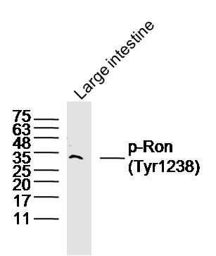
Autophosphorylated in response to ligand binding on Tyr-1238 and Tyr-1239 in the kinase domain leading to further phosphorylation of Tyr-1353 and Tyr-172 in the SLCterminal multifunctional docking site.
Ubiquitinated. Ubiquitination by CBL regulates the receptor stability and activity through proteasomal degradation.

Similarity:
Belongs to the protein kinase superfamily. Tyr protein kinase family.
Contains 3 IPT/TIG domains.
Contains 1 protein kinase domain.
Contains 1 Sema domain.

SWISS:
Q62190

Gene ID:
19882

Database links:

Entrez Gene: 4486 Human

Entrez Gene: 19882 Mouse

Entrez Gene: 300999 Rat

Omim: 600168 Human

SwissProt: Q04912 Human

SwissProt: Q62190 Mouse

Unigene: 517973 Human

Unigene: 3901 Mouse

Unigene: 218659 Rat


Picture

Blank control (Black line): A431 (Black).
Primary Antibody (green line): Rabbit Anti-phospho-Ron(Ser1349) antibody (SL5489R)
Dilution: 3μg /10^6 cells;
Isotype Control Antibody (orange line): Rabbit IgG .
Secondary Antibody (white blue line): Goat anti-rabbit IgG-AF647
Dilution: 3μg /test.
Protocol
The cells were fixed with 4% PFA (10min at room temperature)and then permeabilized with 90% ice-cold methanol for 20 min at room temperature. The cells were then incubated in 5%BSA to block non-specific protein-protein interactions for 30 min at room temperature .Cells stained with Primary Antibody for 30 min at room temperature. The secondary antibody used for 40 min at room temperature. Acquisition of 20,000 events was performed.
Product Feedback Wall
Message :
Your Email :
Copyright © 2007-2018 Sunlong Medical All Rights Reserved.