CD109 is a a novel member of the alpha2-macroglobulin (alpha2M)/complement superfamily. It is a glycosylphosphatidylinositol (GPI)-linked cell surface glycoprotein of approximately 170 kd found on
endothelial cells, activated platelets and T-cells and on a subset of hematopoietic stem and progenitor cells. Although it has been suggested that T-cell CD109 may play a role in antibody-inducing T-helper function and it is known that platelet CD109 carries the Gov alloantigen system, the role of CD109 in hematopoietic cells remains largely unknown.
CD109 has been identified as part of the TGF-beta receptor system in human keratinocytes and upregulation of CD109 expression has been observed in several different types of tumour.
Function:
Modulates negatively TGFB1 signaling in keratinocytes.
Subunit:
Heterodimer; disulfide-linked. Interacts with TGFB1 and TGFBR1. Forms a heteromeric complex with TGFBR1, TGFBR2 and TGFBR3 in a ligand-independent manner.
Subcellular Location:
Cell membrane; Lipid-anchor, GPI-anchor.
Tissue Specificity:
Widely expressed with high level in uterus, aorta, heart, lung, trachea, placenta and in fetal heart, kidney, liver, spleen and lung. Expressed by CD34(+) acute myeloid leukemia cell lines, T-cell lines, activated T-lymphoblasts, endothelial cells and activated platelets. Isoform 5 is expressed in placenta. Isoform 1 is expressed in keratinocytes and placenta.
Post-translational modifications:
N-glycosylated.
2 forms of 150 (p150) and 120 kDa (p120) exist due to proteolytic degradation from a 36 kDa form.
Similarity:
Belongs to the protease inhibitor I39 (alpha-2-macroglobulin) family.
SWISS:
Q6YHK3
Gene ID:
135228
Database links:
Entrez Gene: 135228 Human
Entrez Gene: 235505 Mouse
Entrez Gene: 363104 Rat
Omim: 608859 Human
SwissProt: Q6YHK3 Human
SwissProt: Q8R422 Mouse
Unigene: 399891 Human
Unigene: 32955 Mouse
姬:8个糖基化位点;2 forms of 150 (p150) and 120 kDa (p120) exist due to proteolytic degradation from a 36 kDa form.
| Picture |
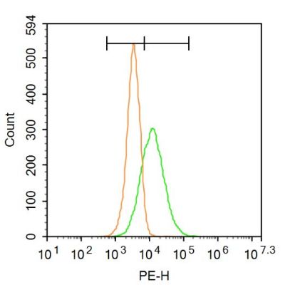
Blank control: A549.
Primary Antibody (green line): Rabbit Anti-CD109 antibody (SL6067R)
Dilution: 3μg /10^6 cells;
Isotype Control Antibody (orange line): Rabbit IgG .
Secondary Antibody : Goat anti-rabbit IgG-PE
Dilution: 3μg /test.
Protocol
The cells were incubated in 5%BSA to block non-specific protein-protein interactions for 30 min at at room temperature .Cells stained with Primary Antibody for 30 min at room temperature. The secondary antibody used for 40 min at room temperature. Acquisition of 20,000 events was performed.
|
|
|