F161A_HUMAN; Fam161a; Family with sequence similarity 161, member A; FLJ13305; Hypothetical protein LOC84140; MGC129982; MGC129983; OTTHUMP00000201353; Protein FAM161A.
Cat:
SL8216R
Species Reactivity:
Human,(predicted: Mouse,Rat,Dog,)
Immunogen:
KLH conjugated synthetic peptide derived from human FAM161A:301-400/660
Format:
Liquid
Storage instructions:
Shipped at 4℃. Store at -20 °C for one year. Avoid repeated freeze/thaw cycles.
Concentration:
1mg/ml
Clonality:
Polyclonal
Isotype:
IgG
Applications:
WB=1:500-2000ELISA=1:5000-10000not yet tested in other applications.optimal dilutions/concentrations should be determined by the end user.
Host:
Rabbit
Product Overview:
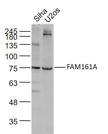
Sample: Siha(Human) Cell Lysate at 30 ugU2os(Human) Cell Lysate at 30 ugPrimary: Anti-FAM161A (SL8216R) at 1/1000 dilutionSecondary: IRDye800CW Goat Anti-Rabbit IgG at 1/20000 dilutionPredicted band size: 77 kDObserved band size: 77 kD
Isoform 1 and isoform 3 are widely expressed with highest levels in retina and testis, with isoform 1 being the mot abundant in all tissues tested.
Involvement in disease:Defects in FAM161A are the cause of retinitis pigmentosa type 28 (RP28) . A retinal dystrophy belonging to the group of pigmentary retinopathies. Retinitis pigmentosa is characterized by retinal pigment deposits visible on fundus examination and primary loss of rod photoreceptor cells followed by secondary loss of cone photoreceptors. Patients typically have night vision blindness and loss of midperipheral visual field. As their condition progresses, they lose their far peripheral visual field and eventually central vision as well.
Function: Involved in ciliogenesis.
Subunit: Interacts (via SLCterminus) with microtubules. Interacts with LCA5, CEP290 and SDCCAG8. Interacts with FAM161B.
Subcellular Location: Cytoplasm, cytoskeleton, cilium basal body. Cell projection, cilium. Note=Localized to the region between the outer and inner photoreceptor segments, corresponding to the photoreceptor connecting cilium.
Tissue Specificity: Isoform 1 and isoform 3 are widely expressed with highest levels in retina and testis, with isoform 1 being the most abundant in all tissues tested.
DISEASE: Defects in FAM161A are the cause of retinitis pigmentosa type 28 (RP28) [MIM:606068]. A retinal dystrophy belonging to the group of pigmentary retinopathies. Retinitis pigmentosa is characterized by retinal pigment deposits visible on fundus examination and primary loss of rod photoreceptor cells followed by secondary loss of cone photoreceptors. Patients typically have night vision blindness and loss of midperipheral visual field. As their condition progresses, they lose their far peripheral visual field and eventually central vision as well.
Sample:
Siha(Human) Cell Lysate at 30 ug
U2os(Human) Cell Lysate at 30 ug
Primary: Anti-FAM161A (SL8216R) at 1/1000 dilution
Secondary: IRDye800CW Goat Anti-Rabbit IgG at 1/20000 dilution
Predicted band size: 77 kD
Observed band size: 77 kD