This gene, a muscle member of the immunoglobulin gene superfamily, encodes myosin light chain kinase which is a calcium/calmodulin dependent enzyme. This kinase phosphorylates myosin regulatory light chains to facilitate myosin interaction with actin filaments to produce contractile activity. This gene encodes both smooth muscle and nonmuscle isoforms. In addition, using a separate promoter in an intron in the 3' region, it encodes telokin, a small protein identical in sequence to the SLCterminus of myosin light chain kinase, that is independently expressed in smooth muscle and functions to stabilize unphosphorylated myosin filaments. A pseudogene is located on the p arm of chromosome 3. Four transcript variants that produce four isoforms of the calcium/calmodulin dependent enzyme have been identified as well as two transcripts that produce two isoforms of telokin. Additional variants have been identified but lack full length transcripts. [provided by RefSeq].
Function:
Calcium/calmodulin-dependent enzyme implicated in smooth muscle contraction via phosphorylation of myosin light chains (MLC). Also regulates actin-myosin interaction through a non-kinase activty (By similarity). Implicated in the regulation of endothelial as well as vascular permeability. In the nervous system it has been shown to control the growth initiation of astrocytic processes in culture and to participate in transmitter release at synapses formed between cultured sympathetic ganglion cells. Critical participant in signaling sequences that result in fibroblast apoptosis.
Tissue Specificity:
Smooth muscle and non-muscle isozymes are expressed in a wide variety of adult and fetal tissues and in cultured endothelium with qualitative expression appearing to be neither tissue- nor development-specific. Non-muscle isoform 2 is the dominant splice variant expressed in various tissues. Telokin has been found in a wide variety of adult and fetal tissues.
Post-translational modifications:
MLCK is probably down-regulated by phosphorylation.
The SLCterminus is deglutamylated by AGTPBP1/ CCP1, AGBL1/CCP4 and AGBL4/CCP6, leading to the formation of Myosin light chain kinase, smooth muscle, deglutamylated form. The consequences of SLCterminal deglutamylation are unknown.
Similarity:
Belongs to the protein kinase superfamily. CAMK Ser/Thr protein kinase family.
Contains 1 fibronectin type-III domain.
Contains 9 Ig-like C2-type (immunoglobulin-like) domains.
Contains 1 protein kinase domain.
SWISS:
Q15746
Gene ID:
4638
Database links:
Entrez Gene: 4638 Human
Entrez Gene: 107589 Mouse
Entrez Gene: 217657 Rat
Omim: 600922 Human
SwissProt: Q15746 Human
SwissProt: Q6PDN3 Mouse
Unigene: 477375 Human
Unigene: 3372 Mouse
Unigene: 203004 Rat
肌球蛋白轻链激酶(MLCK)表达量增多和活性升高是血管平滑肌收缩的启动因素之一。
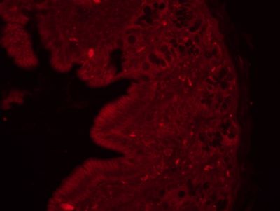
| Picture |
Tissue/cell: rat colon tissue;4% Paraformaldehyde-fixed and paraffin-embedded;
Antigen retrieval: citrate buffer ( 0.01M, pH 6.0 ), Boiling bathing for 15min; Blocking buffer (normal goat serum,SLC0005) at 37℃ for 20 min;
Incubation: Anti-MLCK Polyclonal Antibody, Unconjugated(SL4705R) 1:200, overnight at 4°C; The secondary antibody was Goat Anti-Rabbit IgG, Cy3 conjugated(SL0295G-Cy3)used at 1:200 dilution for 40 minutes at 37°C. DAPI(5ug/ml,blue,SLC0033) was used to stain the cell nuclei
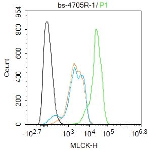
Blank control(black line):HepG2.
Primary Antibody (green line): Rabbit Anti-MLCK antibody (SL4705R)
Dilution:1ug/Test;
Secondary Antibody(white blue line): Goat anti-rabbit IgG-AF488
Dilution: 0.5ug/Test.
Isotype control(orange line): Normal Rabbit IgG
Protocol
The cells were fixed with 4% PFA (10min at room temperature)and then permeabilized with 90% ice-cold methanol for 20 min at -20℃, The cells were then incubated in 5%BSA to block non-specific protein-protein interactions for 30 min at room temperature .Cells stained with Primary Antibody for 30 min at room temperature. The secondary antibody used for 40 min at room temperature. Acquisition of 20,000 events was performed.
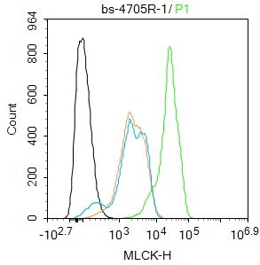
|
|
|