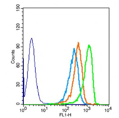
The blue histogram is unstained cells(HepG 2).
The Wathet Blue histogram is cells stained with secondary antibody alone.The Orange histogram is cells stained with rabbit IgG isotype control antibody plus secondary antibody.
The green histogram is cells stained with Rabbit Anti-DOK5 antibody (SL8587R) plus secondary antibody.5μg in 100μL 1 X PBS containing 0.5% BSA.