Home > Product > Antibody > Rabbit Anti-FENS1 antibody
FENS 1; FENS-1; KIAA1435; Phosphoinositide binding protein 1; Phosphoinositide binding protein SR1; Phosphoinositide-binding protein 1; WD repeat and FYVE domain containing 1; WD repeat and FYVE domain-containing protein 1; WD40 and FYVE domain containing
Cat:
SL13169R
Species Reactivity:
Human,Mouse,Rat,(predicted: Dog,Pig,Cow,Horse,Rabbit,)
Immunogen:
KLH conjugated synthetic peptide derived from human FENS1:21-120/410
Format:
Liquid
Storage instructions:
Shipped at 4℃. Store at -20 °C for one year. Avoid repeated freeze/thaw cycles.
Concentration:
1mg/ml
Clonality:
Polyclonal
Isotype:
IgG
Applications:
WB=1:500-2000ELISA=1:5000-10000IHC-P=1:100-500IHC-F=1:100-500ICC=1:100-500IF=1:100-500(Paraffin sections need to do antigen repair)not yet tested in other applications.optimal dilutions/concentrations should be determined by the end user.
Host:
Rabbit
Product Overview:
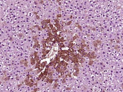
Sample: Lane 1: Cerebrum (Mouse) Tissue Lysate at 40 ugLane 2: Cerebrum (Rat) Tissue Lysate at 40 ugLane 3: Skin (Rat) Tissue Lysate at 40 ugLane 4: Huvec (Human) Cell Lysate at 30 ugLane 5: HepG2 (Human) Cell Lysate at 30 ugLane 6: THP-1 (Human) Cell Lysate at 30 ugPrimary: Anti-FENS1 (SL13169R) at 1/1000 dilutionSecondary: IRDye800CW Goat Anti-Rabbit IgG at 1/20000 dilutionPredicted band size: 46 kDObserved band size: 48 kDSample: Lane 1: U87MG (Human) Cell Lysate at 30 ugLane 2: HepG2 (Human) Cell Lysate at 30 ugLane 3: U2os (Human) Cell Lysate at 30 ugLane 4: A431 (Human) Cell Lysate at 30 ugPrimary: Anti-FENS1 (SL13169R) at 1/1000 dilutionSecondary: IRDye800CW Goat Anti-Rabbit IgG at 1/20000 dilutionPredicted band size: 46 kDObserved band size: 46 kD Paraformaldehyde-fixed, paraffin embedded (Rat liver); Antigen retrieval by boiling in sodium citrate buffer (pH6.0) for 15min; Block endogenous peroxidase by 3% hydrogen peroxide for 20 minutes; Blocking buffer (normal goat serum) at 37°C for 30min; Antibody incubation with (FENS1) Polyclonal Antibody, Unconjugated (SL13169R) at 1:400 overnight at 4°C, followed by operating according to SP Kit(Rabbit) (sp-0023) instructionsand DAB staining.
More
Unit:
Price: $
Product PDFs
Datasheet:


WD-repeats are motifs that are found in a variety of proteins and are characterized by a conserved core of 40-60 amino acids that commonly form a tertiary propeller structure. While proteins that contain WD-repeats participate in a wide range of cellular functions, they are generally involved in regulatory mechanisms concerning chromatin assembly, cell cycle control, signal transduction, RNA processing, apoptosis and vesicular trafficking. WDFY1 (WD repeat and FYVE domain containing 1), also known as WDF1, FENS-1 or ZFYVE17, is a 410 amino acid protein that localizes to the early endosome and contains one FYVE-type zinc finger and seven WD repeats through which it may play a role in protein trafficking and signal transduction.

Subunit:
Binds PtdIns3P in vitro with high specificity over other phosphoinositides.

Subcellular Location:
Early endosome.

Similarity:
Contains 1 FYVE-type zinc finger.
Contains 7 WD repeats.

SWISS:
Q8IWB7

Gene ID:
57590

Database links:

Entrez Gene: 57590 Human

Entrez Gene: 69368 Mouse

Entrez Gene: 301549 Rat

SwissProt: Q8IWB7 Human

Unigene: 368359 Human

Unigene: 8557 Rat


Picture

Sample:
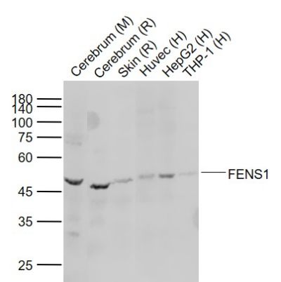
Lane 1: Cerebrum (Mouse) Tissue Lysate at 40 ug
Lane 2: Cerebrum (Rat) Tissue Lysate at 40 ug
Lane 3: Skin (Rat) Tissue Lysate at 40 ug
Lane 4: Huvec (Human) Cell Lysate at 30 ug
Lane 5: HepG2 (Human) Cell Lysate at 30 ug
Lane 6: THP-1 (Human) Cell Lysate at 30 ug
Primary: Anti-FENS1 (SL13169R) at 1/1000 dilution
Secondary: IRDye800CW Goat Anti-Rabbit IgG at 1/20000 dilution
Predicted band size: 46 kD
Observed band size: 48 kD
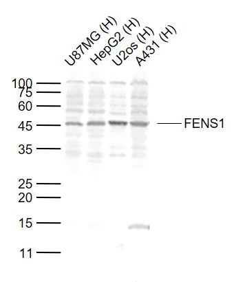
Sample:
Lane 1: U87MG (Human) Cell Lysate at 30 ug
Lane 2: HepG2 (Human) Cell Lysate at 30 ug
Lane 3: U2os (Human) Cell Lysate at 30 ug
Lane 4: A431 (Human) Cell Lysate at 30 ug
Primary:
Anti-FENS1 (SL13169R) at 1/1000 dilution
Secondary: IRDye800CW Goat Anti-Rabbit IgG at 1/20000 dilution
Predicted band size: 46 kD
Observed band size: 46 kD
Paraformaldehyde-fixed, paraffin embedded (Rat liver); Antigen retrieval by boiling in sodium citrate buffer (pH6.0) for 15min; Block endogenous peroxidase by 3% hydrogen peroxide for 20 minutes; Blocking buffer (normal goat serum) at 37°C for 30min; Antibody incubation with (FENS1) Polyclonal Antibody, Unconjugated (SL13169R) at 1:400 overnight at 4°C, followed by operating according to SP Kit(Rabbit) (sp-0023) instructionsand DAB staining.
Product Feedback Wall
Message :
Your Email :
Copyright © 2007-2018 Sunlong Medical All Rights Reserved.