MAGE-H1 is a 219 amino acid protein that contains a type II MAGE homology domain (MHD). Enhanced ligand stimulation promotes MAGE-H1 interaction with the type II death domain of NGFR p75. It is suggested that MAGE-H1 accelerates differentiation in response to nerve growth factor in cells.
Function:
Acts as a scaffolding protein at cell-cell junctions, thereby regulating various cellular and signaling processes. Cooperates with PTEN to modulate the kinase activity of AKT1. Its interaction with PTPRB and tyrosine phosphorylated proteins suggests that it may link receptor tyrosine phosphatase with its substrates at the plasma membrane. In polarized epithelial cells, involved in efficient trafficking of TGFA to the cell surface. Regulates the ability of LPAR2 to activate ERK and RhoA pathways. Regulates the JNK signaling cascade via its interaction with FZD4 and VANGL2.
Subcellular Location:
Cell membrane. Cell junction > tight junction. Nucleus. Concentrates in specific sites at the plasma membrane and in the nucleus. In epithelial cells, it localizes at tight junctions.
Tissue Specificity:
Widely expressed.
Post-translational modifications:
Ubiquitinated following interaction with HPV E6 protein, leading to its degradation by the proteasome. Degradation is independent of E6AP ubiquitin ligase complex.
Similarity:
Belongs to the MAGUK family.
Contains 1 guanylate kinase-like domain.
Contains 6 PDZ (DHR) domains.
Contains 2 WW domains.
SWISS:
Q5TCQ9
Gene ID:
260425
Database links:
Entrez Gene: 260425 Human
Entrez Gene: 99470 Mouse
Entrez Gene: 245903 Rat
SwissProt: Q5TCQ9 Human
SwissProt: Q9EQJ9 Mouse
SwissProt: Q9JK71 Rat
Unigene: 486189 Human
Unigene: 264849 Mouse
Unigene: 162623 Rat
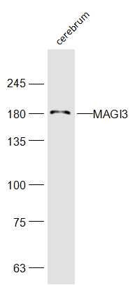
| Picture |
Sample:
Cerebrum(Mouse) Cell Lysate at 40 ug
Primary: Anti-MAGI3(SL18629R) at 1/300 dilution
Secondary: IRDye800CW Goat Anti-Rabbit IgG at 1/20000 dilution
Predicted band size: 166kD
Observed band size: 36kD
|
|
|