8030489C12Rik;
Cell migration inducing 1;
Cell migration-inducing gene 1 protein;
MGC116271;
MIG1;
Protein SKD1;
Skd1;
SKD1B;
Suppressor of K(+) transport growth defect 1;
Suppressor of K+ transport defect 1;
Vacuolar protein sorting 4 homolog B
Cat:
SL12778R
Species Reactivity:
Human,Mouse,(predicted: Rat,Pig,Cow,)
Immunogen:
KLH conjugated synthetic peptide derived from human VPS4B:301-400/444
Format:
Liquid
Storage instructions:
Shipped at 4℃. Store at -20 °C for one year. Avoid repeated freeze/thaw cycles.
Concentration:
1mg/ml
Clonality:
Polyclonal
Isotype:
IgG
Applications:
WB=1:500-2000ELISA=1:5000-10000IHC-P=1:100-500IHC-F=1:100-500ICC=1:100-500IF=1:100-500(Paraffin sections need to do antigen repair)not yet tested in other applications.optimal dilutions/concentrations should be determined by the end user.
Host:
Rabbit
Product Overview:
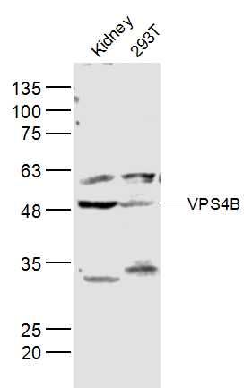
Sample: Kidney (Mouse) Lysate at 40 ug293T(Human) Cell Lysate at 40 ugPrimary: Anti-VPS4B (SL12778R) at 1/300 dilutionSecondary: IRDye800CW Goat Anti-Rabbit IgG at 1/20000 dilutionPredicted band size: 49 kDObserved band size: 49 kD
The protein encoded by this gene is a member of the AAA protein family (ATPases associated with diverse cellular activities), and is the homolog of the yeast Vps4 protein. In humans, two paralogs of the yeast protein have been identified. The former share a high degree of aa sequence similarity with each other, and also with yeast Vps4 and mouse Skd1 proteins. Mouse Skd1 (suppressor of K+ transport defect 1) has been shown to be a yeast Vps4 ortholog. Functional studies indicate that both human paralogs associate with the endosomal compartments, and are involved in intracellular protein trafficking, similar to Vps4 protein in yeast. The gene encoding this paralog has been mapped to chromosome 18; the gene for the other resides on chromosome 16. [provided by RefSeq, Jul 2008]
Function: Involved in late steps of the endosomal multivesicular bodies (MVB) pathway. Recognizes membrane-associated ESCRT-III assemblies and catalyzes their disassembly, possibly in combination with membrane fission. Redistributes the ESCRT-III components to the cytoplasm for further rounds of MVB sorting. MVBs contain intraluminal vesicles (ILVs) that are generated by invagination and scission from the limiting membrane of the endosome and mostly are delivered to lysosomes enabling degradation of membrane proteins, such as stimulated growth factor receptors, lysosomal enzymes and lipids. In conjunction with the ESCRT machinery also appears to function in topologically equivalent membrane fission events, such as the terminal stages of cytokinesis and enveloped virus budding (HISLV1 and other lentiviruses).
Subcellular Location: Prevacuolar compartment membrane. Late endosome membrane. Membrane-associated in the prevacuolar endosomal compartment. Localized in HISLV1 particles purified from acutely infected cells.
Tissue Specificity: Ubiquitously expressed.
Post-translational modifications: Phosphorylated upon DNA damage, probably by ATM or ATR.
Similarity: Belongs to the AAA ATPase family.
Contains 1 MIT domain.
Sample:
Kidney (Mouse) Lysate at 40 ug
293T(Human) Cell Lysate at 40 ug
Primary: Anti-VPS4B (SL12778R) at 1/300 dilution
Secondary: IRDye800CW Goat Anti-Rabbit IgG at 1/20000 dilution
Predicted band size: 49 kD
Observed band size: 49 kD
Product Feedback Wall
Specific References (1) | SL12778R has been referenced in 1 publications.
[IF=3.298] Chen Y et al. The role of infectious hematopoietic necrosis virus (IHNV) proteins in recruiting the ESCRT pathway through three ways in the host cells of fish during IHNV budding. Fish Shellfish Immunol. 2019 Jul 9;92:833-841. WB ; Chinook.