Home > Product > Antibody > Rabbit Anti-TGF-beta 3 antibody
Transforming growth factor beta-3 proprotein; TGFB3; ARVD; FLJ16571; MGC105479; MGC118722; TGF beta 3; TGF beta3; TGF-beta-3; TGF-beta 3; TGFB 3; Transforming growth factor beta-3; TGFB3_HUMAN.
Cat:
SL20413R
Species Reactivity:
Human,Mouse,(predicted: Rat,)
Immunogen:
KLH conjugated synthetic peptide derived from human TGF-beta 3:321-412/412
Format:
Liquid
Storage instructions:
Shipped at 4℃. Store at -20 °C for one year. Avoid repeated freeze/thaw cycles.
Concentration:
1mg/ml
Clonality:
Polyclonal
Isotype:
IgG
Applications:
WB=1:500-2000ELISA=1:5000-10000not yet tested in other applications.optimal dilutions/concentrations should be determined by the end user.
Host:
Rabbit
Product Overview:
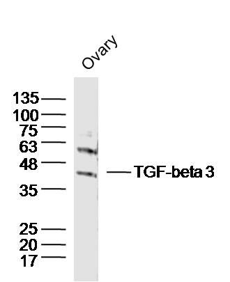
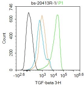
Sample:Ovary (Mouse)Lysate at 40 ugPrimary: Anti-TGF-beta 3(SL20413R)at 1/300 dilutionSecondary: IRDye800CW Goat Anti-RabbitIgG at 1/20000 dilutionPredicted band size: 30'47kDObserved band size: 47kDBlank control(black line):SH-SY5Y. Primary Antibody (green line): Rabbit Anti-TGF-beta 3 antibody (SL20413R) Dilution:1ug/Test; Secondary Antibody : Goat anti-rabbit IgG-AF488Dilution: 0.5ug/Test. Negative control(white blue line): PBSIsotype control(orange line): Normal Rabbit IgGProtocolThe cells were fixed with 4% PFA (10min at room temperature)and then permeabilized with 90% ice-cold methanol for 20 min at -20℃.The cells were then incubated in 5%BSA to block non-specific protein-protein interactions for 30 min at room temperature .Cells stained with Primary Antibody for 30 min at room temperature. The secondary antibody used for 40 min at room temperature. Acquisition of 20,000 events was performed.
More
Unit:
Price: $
Product PDFs
Datasheet:


The TGFBeta superfamily is composed of numerous growth and differentiation factors, including transforming growth factors Beta(TGFBeta) 1,2 and 3; placental transforming growth factor (PTGFBeta);growth /differentiation factors (GDFs); mullerian inhibiting substance (MIS);bone morphogenic proteins (BMPs);glial cell line-derived neurotrophic factor (GDNF);inhibins or activins (Alpha,Beta-A, Beta-B and Beta-C),Lefty and Nodal. Members of the TGF superfamily are involved in embryonic development and adult tissue homeostasis.

Function:
Involved in embryogenesis and cell differentiation.

Subunit:
Homodimer; disulfide-linked. Interacts with ASPN.

Subcellular Location:
Secreted.

DISEASE:
Defects in TGFB3 are a cause of familial arrhythmogenic right ventricular dysplasia type 1 (ARVD1) [MIM:107970]; also known as arrhythmogenic right ventricular cardiomyopathy 1 (ARVC1). ARVD is an autosomal dominant disease characterized by partial degeneration of the myocardium of the right ventricle, electrical instability, and sudden death. It is clinically defined by electrocardiographic and angiographic criteria; pathologic findings, replacement of ventricular myocardium with fatty and fibrous elements, preferentially involve the right ventricular free wall.

Similarity:
Belongs to the TGF-beta family.

SWISS:
P10600

Gene ID:
7043

Database links:

Entrez Gene: 7043 Human

Entrez Gene: 2369 Mouse

Entrez Gene: 25717 Rat

Omim: 190230 Human

SwissProt: P10600 Human

SwissProt: P17125 Mouse

SwissProt: Q07258 Rat

Unigene: 592317 Human

Unigene: 3992 Mouse

Unigene: 7018 Rat


生长因子和激素( Growth Factor and Hormones) TGF是一种多效生长因子,对上皮细胞增值有潜在抑制作用,可抑制肿瘤生长,用于许多恶性肿瘤如:胃癌、肺癌、膀胱癌、肾癌、前列腺癌、结肠癌等多种恶性肿瘤的研究。分子量:47kDa。此抗体是TGFβ3 特异性抗体,与TGF-β1和TGF-β2没有交叉反应。 Picture

Sample:Ovary (Mouse)Lysate at 40 ug
Primary: Anti-TGF-beta 3(SL20413R)at 1/300 dilution
Secondary: IRDye800CW Goat Anti-RabbitIgG at 1/20000 dilution
Predicted band size: 30'47kD
Observed band size: 47kD
Blank control(black line):SH-SY5Y.
Primary Antibody (green line): Rabbit Anti-TGF-beta 3 antibody (SL20413R)
Dilution:1ug/Test;
Secondary Antibody : Goat anti-rabbit IgG-AF488
Dilution: 0.5ug/Test.
Negative control(white blue line): PBS
Isotype control(orange line): Normal Rabbit IgG
Protocol
The cells were fixed with 4% PFA (10min at room temperature)and then permeabilized with 90% ice-cold methanol for 20 min at -20℃.The cells were then incubated in 5%BSA to block non-specific protein-protein interactions for 30 min at room temperature .Cells stained with Primary Antibody for 30 min at room temperature. The secondary antibody used for 40 min at room temperature. Acquisition of 20,000 events was performed.
Product Feedback Wall
Message :
Your Email :
Copyright © 2007-2018 Sunlong Medical All Rights Reserved.