Home > Product > Antibody > Rabbit Anti-ECT2 antibody
ECT 2; ECT2; ECT-2; ECT2 protein; Epithelial cell transforming sequence 2; Epithelial cell transforming sequence 2 oncogene; Epithelial cell transforming sequence 2 oncogene protein; FLJ10461; MGC138291; Protein ECT2; ECT2_HUMAN; Epithelial cell-transform
Cat:
SL23904R
Species Reactivity:
Rat,(predicted: Human,Mouse,Dog,Pig,Rabbit,Sheep,)
Immunogen:
KLH conjugated synthetic peptide derived from human ECT2 :751-850/914
Format:
Liquid
Storage instructions:
Shipped at 4℃. Store at -20 °C for one year. Avoid repeated freeze/thaw cycles.
Concentration:
1mg/1ml
Clonality:
Polyclonal
Isotype:
IgG
Applications:
WB=1:500-2000IHC-P=1:100-500IHC-F=1:100-500ICC=1:100-500IF=1:100-500(Paraffin sections need to do antigen repair)not yet tested in other applications.optimal dilutions/concentrations should be determined by the end user.
Host:
Rabbit
Product Overview:
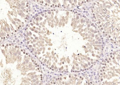
Paraformaldehyde-fixed, paraffin embedded (rat testis tissue); Antigen retrieval by boiling in sodium citrate buffer (pH6.0) for 15min; Block endogenous peroxidase by 3% hydrogen peroxide for 20 minutes; Blocking buffer (normal goat serum) at 37°C for 30min; Antibody incubation with (ECT2) Polyclonal Antibody, Unconjugated (SL23904R) at 1:400 overnight at 4°C, followed by operating according to SP Kit(Rabbit) (sp-0023) instructionsand DAB staining.
More
Unit:
Price: $
Product PDFs
Datasheet:


The protein encoded by this gene is a transforming protein that is related to Rho-specific exchange factors and yeast cell cycle regulators. The expression of this gene is elevated with the onset of DNA synthesis and remains elevated during G2 and M phases. In situ hybridization analysis showed that expression is at a high level in cells undergoing mitosis in regenerating liver. Thus, this protein is expressed in a cell cycle-dependent manner during liver regeneration, and is thought to have an important role in the regulation of cytokinesis. [provided by RefSeq].

Function:
Guanine nucleotide exchange factor (GEF) that catalyzes the exchange of GDP for GTP. Promotes guanine nucleotide exchange on the Rho family members of small GTPases, like RHOA, RHOC, RAC1 and CDC42. Required for signal transduction pathways involved in the regulation of cytokinesis. Component of the centralspindlin complex that serves as a microtubule-dependent and Rho-mediated signaling required for the myosin contractile ring formation during the cell cycle cytokinesis. Regulates the translocation of RHOA from the central spindle to the equatorial region. Plays a role in the control of mitotic spindle assembly; regulates the activation of CDC42 in metaphase for the process of spindle fibers attachment to kinetochores before chromosome congression. Involved in the regulation of epithelial cell polarity; participates in the formation of epithelial tight junctions in a polarity complex PARD3-PARD6-protein kinase PRKCQ-dependent manner. Plays a role in the regulation of neurite outgrowth. Inhibits Phenobarbital (PB)-induced NR1I3 nuclear translocation. Stimulates the activity of RAC1 through its association with the oncogenic PARD6A-PRKCI complex in cancer cells, thereby acting to coordinately drive tumor cell proliferation and invasion. Also stimulates genotoxic stress-induced RHOB activity in breast cancer cells leading to their cell death.

Subunit:
Interacts with NR1I3 (By similarity). Homodimer. Homooligomer. Found in the centralspindlin complex. Interacts (Thr-359 phosphorylated form) with PARD6A; the interaction is observed in cancer cells. Interacts (Thr-359 phosphorylated form) with PRKCI; the interaction is observed in cancer cells. Interacts with PKP4; the interaction is observed at the midbody. Interacts with RACGAP1; the interaction is direct, occurs in a microtubule-dependent manner, is inhibited in metaphase by phosphorylation of ECT2 on Thr-373 and is stimulated in early anaphase by dephosphorylation of ECT2 probably on Thr-373 through CDK1 activity. Interacts with PLK1; the interaction is stimulated upon its phosphorylation on Thr-444. Associates with RACGAP1 at anaphase and during cytokinesis. Interacts with KIF23, PARD3, PARD6A, PARD6B and PRKCQ.

Subcellular Location:
Nucleus. Cytoplasm. Cytoplasm, cytoskeleton, spindle. Cleavage furrow. Midbody. Cell junction. Cell junction, tight junction. Note=Sequestered within the nucleus during interphase. Dispersed throughout the cytoplasm upon breakdown of the nuclear envelope during mitosis. Colocalizes with the centralspindlin complex to the mitotic spindles during anaphase/metaphase, the cleavage furrow during telophase and at the midbody at the end of cytokinesis. Colocalized with RhoA at the midbody. Its subcellular localization to tight junction is increased by calcium. Localized predominantly in the cytoplasm of numerous carcinoma cells.

Tissue Specificity:
Expressed in lung epithelial cells (at protein level). Expressed in squamous cell carcinoma, primary non-small cell lung cancer tumors and lung adenocarcinoma. [INDUCTION] Up-regulated by calcium in cells forming cell-cell contact sites. Up-regulated by DNA damaging agents like H(2)O(2) or ionizing radiation (IR).

Post-translational modifications:
Phosphorylated by PLK1 in vitro. Hyperphosphorylated during the G2 phase of the cell cycle. Phosphorylation at Thr-373 occurs during the G2/M phase, relieves its auto-inhibition status and stimulates its GEF activity. Phosphorylation at Thr-444 in G2/M phase is required for subsequent binding with PLK1 and Rho exchange activation. Dephosphorylated at the time of cytokinesis. Phosphorylation at Thr-359 is required for its transformation activity in cancer cells.

Similarity:
Contains 2 BRCT domains.
Contains 1 DH (DBL-homology) domain.
Contains 1 PH domain.

SWISS:
Q9H8V3

Gene ID:
1894

Database links:

Entrez Gene: 1894 Human

Entrez Gene: 1725 Mouse

Entrez Gene: 361921 Rat

Omim: 600586 Human

SwissProt: Q9H8V3 Human

SwissProt: Q07139 Mouse

Unigene: 261453 Mouse

Unigene: 168292 Rat


Picture

Paraformaldehyde-fixed, paraffin embedded (rat testis tissue); Antigen retrieval by boiling in sodium citrate buffer (pH6.0) for 15min; Block endogenous peroxidase by 3% hydrogen peroxide for 20 minutes; Blocking buffer (normal goat serum) at 37°C for 30min; Antibody incubation with (ECT2) Polyclonal Antibody, Unconjugated (SL23904R) at 1:400 overnight at 4°C, followed by operating according to SP Kit(Rabbit) (sp-0023) instructionsand DAB staining.
Product Feedback Wall
Message :
Your Email :
Copyright © 2007-2018 Sunlong Medical All Rights Reserved.