Home > Product > Antibody > Rabbit Anti-phospho-MDM2 (Ser188 + Ser186) antibody
MDM2 (phospho S188 + S186); p-MDM2 (phospho S188 + S186); ACTFS; Double minute 2 protein; E3 ubiquitin-protein ligase Mdm2; Hdm 2; Hdm2; HDMX; MDM 2; MDM2; MDM2 oncogene E3 ubiquitin protein ligase; Mdm2 p53 E3 ubiquitin protein ligase homolog;
Cat:
SL18747R
Species Reactivity:
Human,
Immunogen:
KLH conjugated synthesised phosphopeptide derived from human MDM2 around the phosphorylation site of Ser188 + Ser186:HK(p-S)D(p-S)IS
Format:
Liquid
Storage instructions:
Shipped at 4℃. Store at -20 °C for one year. Avoid repeated freeze/thaw cycles.
Concentration:
1mg/ml
Clonality:
Polyclonal
Isotype:
IgG
Applications:
WB=1:500-2000ELISA=1:5000-10000IHC-P=1:100-500IHC-F=1:100-500Flow-Cyt=1ug/TestICC=1:100IF=1:100-500(Paraffin sections need to do antigen repair)not yet tested in other applications.optimal dilutions/concentrations should be determined by the end user.
Host:
Rabbit
Product Overview:
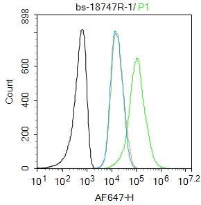
Blank control:Molt4. Primary Antibody (green line): Rabbit Anti-phospho-MDM2 (Ser188 + Ser186) antibody (SL18747R) Dilution: 1μg /10^6 cells; Isotype Control Antibody (orange line): Rabbit IgG . Secondary Antibody : Goat anti-rabbit IgG-AF647Dilution: 1μg /test. ProtocolThe cells were fixed with 4% PFA (10min at room temperature)and then permeabilized with 90% ice-cold methanol for 20 min at-20℃. The cells were then incubated in 5%BSA to block non-specific protein-protein interactions for 30 min at room temperature .Cells stained with Primary Antibody for 30 min at room temperature. The secondary antibody used for 40 min at room temperature. Acquisition of 20,000 events was performed.
More
Unit:
Price: $
Product PDFs
Datasheet:


This gene encodes a nuclear-localized E3 ubiquitin ligase. The encoded protein can promote tumor formation by targeting tumor suppressor proteins, such as p53, for proteasomal degradation. This gene is itself transcriptionally-regulated by p53. Overexpression or amplification of this locus is detected in a variety of different cancers. There is a pseudogene for this gene on chromosome 2. Alternative splicing results in a multitude of transcript variants, many of which may be expressed only in tumor cells. [provided by RefSeq, Jun 2013]

Function:
E3 ubiquitin-protein ligase that mediates ubiquitination of p53/TP53, leading to its degradation by the proteasome. Inhibits p53/TP53- and p73/TP73-mediated cell cycle arrest and apoptosis by binding its transcriptional activation domain. Also acts as an ubiquitin ligase E3 toward itself and ARRB1. Permits the nuclear export of p53/TP53. Promotes proteasome-dependent ubiquitin-independent degradation of retinoblastoma RB1 protein. Inhibits DAXX-mediated apoptosis by inducing its ubiquitination and degradation. Component of the TRIM28/KAP1-MDM2-p53/TP53 complex involved in stabilizing p53/TP53. Also component of the TRIM28/KAP1-ERBB4-MDM2 complex which links growth factor and DNA damage response pathways.

Subcellular Location:
Nucleus > nucleoplasm. Cytoplasm. Nucleus > nucleolus. Expressed predominantly in the nucleoplasm. Interaction with ARF(P14) results in the localization of both proteins to the nucleolus. The nucleolar localization signals in both ARF(P14) and MDM2 may be necessary to allow efficient nucleolar localization of both proteins. Colocalizes with RASSF1 isoform A in the nucleus.

Tissue Specificity:
Ubiquitous. Isoform Mdm2-A, isoform Mdm2-B, isoform Mdm2-C, isoform Mdm2-D, isoform Mdm2-E, isoform Mdm2-F and isoform Mdm2-G are observed in a range of cancers but absent in normal tissues.

Post-translational modifications:
Phosphorylated in response to ionizing radiation in an ATM-dependent manner. Auto-ubiquitinated; which leads to proteasomal degradation. Deubiquitinated by USP2 leads to its accumulation and increases deubiquitinilation and degradation of p53/TP53. Deubiquitinated by USP7; leading to stabilize it.

DISEASE:
Note=Seems to be amplified in certain tumors (including soft tissue sarcomas, osteosarcomas and gliomas). A higher frequency of splice variants lacking p53 binding domain sequences was found in late-stage and high-grade ovarian and bladder carcinomas. Four of the splice variants show loss of p53 binding.

Similarity:
Belongs to the MDM2/MDM4 family.
Contains 1 RanBP2-type zinc finger.
Contains 1 RING-type zinc finger.
Contains 1 SWIB domain.

SWISS:
Q00987

Gene ID:
4193

Database links:

Entrez Gene: 4193 Human

Omim: 164785 Human

SwissProt: Q00987 Human

Unigene: 484551 Human


Picture

Blank control:Molt4.
Primary Antibody (green line): Rabbit Anti-phospho-MDM2 (Ser188 + Ser186) antibody (SL18747R)
Dilution: 1μg /10^6 cells;
Isotype Control Antibody (orange line): Rabbit IgG .
Secondary Antibody : Goat anti-rabbit IgG-AF647
Dilution: 1μg /test.
Protocol
The cells were fixed with 4% PFA (10min at room temperature)and then permeabilized with 90% ice-cold methanol for 20 min at-20℃. The cells were then incubated in 5%BSA to block non-specific protein-protein interactions for 30 min at room temperature .Cells stained with Primary Antibody for 30 min at room temperature. The secondary antibody used for 40 min at room temperature. Acquisition of 20,000 events was performed.
Product Feedback Wall
Message :
Your Email :
Copyright © 2007-2018 Sunlong Medical All Rights Reserved.