Home > Product > Antibody > Rabbit Anti-Aurora A antibody
AIK; ARK-1; ARK1; AURA; AURKA; Aurora 2; Aurora Family Kinase 1; aurora kinase A; Aurora Related Kinase 1; Aurora-related kinase 1; Aurora/IPL1 Like Kinase; Aurora/IPL1 Related Kinase 1; Aurora/IPL1-related kinase 1; AURORA2; AYK1; BRAK; Breast Tumor Ampl
Cat:
SLM52018R
Species Reactivity:
Human,Rat,(predicted: Mouse,)
Immunogen:
Recombinant human Aurora A protein, around C-terminal 250aa
Format:
Liquid
Storage instructions:
Shipped at 4℃. Store at -20 °C for one year. Avoid repeated freeze/thaw cycles.
Concentration:
1mg/ml
Clonality:
Monoclonal
Isotype:
IgG
Applications:
WB=1:500-2000IHC-P=1:100-400IHC-F=1:100-400Flow-Cyt=1:50ICC=1:50IF=1:100-400(Paraffin sections need to do antigen repair)not yet tested in other applications.optimal dilutions/concentrations should be determined by the end user.
Host:
Rabbit
Product Overview:
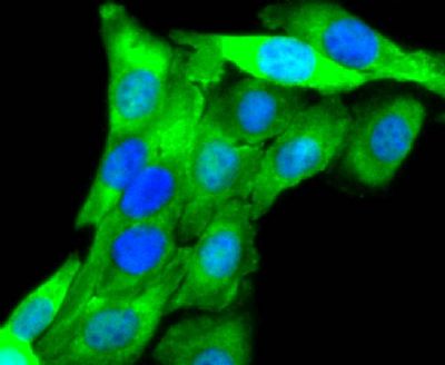
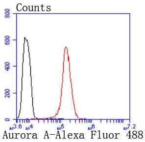
Sample: Lane 1: K562 (Human) Cell Lysate at 30 ugPrimary: Anti-Aurora A (SLM52018R) at 1/500 dilutionSecondary: Goat Anti-Rabbit IgG - HRP at 1/8000 dilutionPredicted band size: 48 kDObserved band size: 46 kDParaformaldehyde-fixed, paraffin embedded (rat testis); Antigen retrieval by boiling in sodium citrate buffer (pH6.0) for 15min; Block endogenous peroxidase by 3% hydrogen peroxide for 20 minutes; Blocking buffer (normal goat serum) at 37°C for 30min; Antibody incubation with (Aurora A) Monoclonal Antibody, Unconjugated (SLM52018R) at 1:50 overnight at 4°C, followed by operating according to SP Kit(Rabbit) (sp-0023) instructionsand DAB staining.Hela cell; 4% Paraformaldehyde-fixed; Triton X-100 at room temperature for 20 min; Blocking buffer (normal goat serum, SLC0005) at 37°C for 20 min; Antibody incubation with (Aurora A) monoclonal Antibody, Unconjugated (SLM52018R) 1:50, 90 minutes at 37°C; followed by a conjugated Goat Anti-Rabbit IgG antibody at 37°C for 90 minutes, DAPI (blue, C02-04002) was used to stain the cell nuclei.NIH/3T3 cell; 4% Paraformaldehyde-fixed; Triton X-100 at room temperature for 20 min; Blocking buffer (normal goat serum, SLC0005) at 37°C for 20 min; Antibody incubation with (Aurora A) monoclonal Antibody, Unconjugated (SLM52018R) 1:50, 90 minutes at 37°C; followed by a conjugated Goat Anti-Rabbit IgG antibody at 37°C for 90 minutes, DAPI (blue, C02-04002) was used to stain the cell nuclei.SKOSLV3 cell; 4% Paraformaldehyde-fixed; Triton X-100 at room temperature for 20 min; Blocking buffer (normal goat serum, SLC0005) at 37°C for 20 min; Antibody incubation with (Aurora A) monoclonal Antibody, Unconjugated (SLM52018R) 1:50, 90 minutes at 37°C; followed by a conjugated Goat Anti-Rabbit IgG antibody at 37°C for 90 minutes, DAPI (blue, C02-04002) was used to stain the cell nuclei.Blank control: Hela. Primary Antibody (green line): Rabbit Anti-Aurora A antibody (SLM52018R) Dilution: 1:50; Isotype Control Antibody (orange line): Rabbit IgG . Secondary Antibody : Goat anti-rabbit IgG-AF488Dilution: 1:1000. ProtocolThe cells were fixed with 4% PFA (10min at room temperature)and then permeabilized with 0.1% PBST for 20 min at room temperature. The cells were then incubated in 5%BSA to block non-specific protein-protein interactions for 30 min at room temperature .Cells stained with Primary Antibody for 30 min at room temperature. The secondary antibody used for 40 min at room temperature. Acquisition of 20,000 events was performed.
More
Unit:
Price: $
Product PDFs
Datasheet:


The protein encoded by this gene is a cell cycle-regulated kinase that appears to be involved in microtubule formation and/or stabilization at the spindle pole during chromosome segregation. The encoded protein is found at the centrosome in interphase cells and at the spindle poles in mitosis. This gene may play a role in tumor development and progression. A processed pseudogene of this gene has been found on chromosome 1, and an unprocessed pseudogene has been found on chromosome 10. Multiple transcript variants encoding the same protein have been found for this gene. [provided by RefSeq, Jul 2008]

Function:
Contributes to the regulation of cell cycle progression. Required for normal mitosis. Associates with the centrosome and the spindle microtubules during mitosis and functions in centrosome maturation, spindle assembly, maintenance of spindle bipolarity, centrosome separation and mitotic checkpoint control. Phosphorylates numerous target proteins, including ARHGEF2, BRCA1, KIF2A, NDEL1, PARD3, PLK1 and BORA. Regulates KIF2A tubulin depolymerase activity. Required for normal axon formation. Plays a role in microtubule remodeling during neurite extension. Important for microtubule formation and/or stabilization.

Subunit:
Interacts with FBXL7. Interacts with CPEB1, JTB, TACC1, TPX2, PPP2CA, as well as with the protein phosphatase type 1 (PP1) isoforms PPP1CA, PPP1CB and PPP1CC. Interacts also with its substrates ARHGEF2, BORA, BRCA1, KIF2A, PARD3, and p53/TP53. Interaction with BORA promotes phosphorylation of PLK1. Interacts with PIFO. Interacts with GADD45A, competing with its oligomerization. Interacts (via SLCterminus) with AUNIP (via SLCterminus). Identified in a complex with AUNIP and NIN. Interacts with FRY; this interaction facilitates AURKA-mediated PLK1 phosphorylation.

Subcellular Location:
Cytoplasm.

Tissue Specificity:
Highly expressed in testis and weakly in skeletal muscle, thymus and spleen. Also highly expressed in colon, ovarian, prostate, neuroblastoma, breast and cervical cancer cell lines.

Post-translational modifications:
Activated by phosphorylation at Thr-288; this brings about a change in the conformation of the activation segment. Phosphorylation at Thr-288 varies during the cell cycle and is highest during M phase. Autophosphorylated at Thr-288 upon TPX2 binding. Phosphorylated upon DNA damage, probably by ATM or ATR. Ubiquitinated by CHFR, leading to its degradation by the proteasome. Ubiquitinated by the anaphase-promoting complex (APC), leading to its degradation by the proteasome.

Similarity:
Belongs to the protein kinase superfamily. Ser/Thr protein kinase family. Aurora subfamily.
Contains 1 protein kinase domain.

SWISS:
O14965

Gene ID:
6790

Database links:

Entrez Gene: 6790 Human

Entrez Gene: 20878 Mouse

Omim: 603072 Human

SwissProt: O14965 Human

SwissProt: P97477 Mouse

Unigene: 250822 Human

Unigene: 249363 Mouse


Picture

Sample:
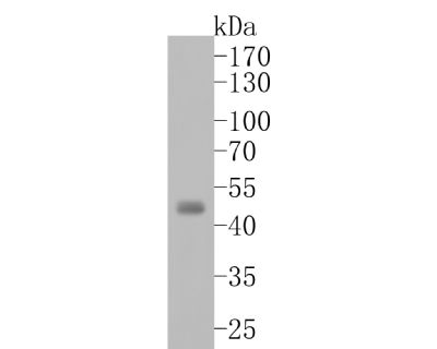
Lane 1: K562 (Human) Cell Lysate at 30 ug
Primary: Anti-Aurora A (SLM52018R) at 1/500 dilution
Secondary: Goat Anti-Rabbit IgG - HRP at 1/8000 dilution
Predicted band size: 48 kD
Observed band size: 46 kD
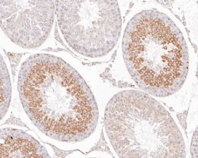
Paraformaldehyde-fixed, paraffin embedded (rat testis); Antigen retrieval by boiling in sodium citrate buffer (pH6.0) for 15min; Block endogenous peroxidase by 3% hydrogen peroxide for 20 minutes; Blocking buffer (normal goat serum) at 37°C for 30min; Antibody incubation with (Aurora A) Monoclonal Antibody, Unconjugated (SLM52018R) at 1:50 overnight at 4°C, followed by operating according to SP Kit(Rabbit) (sp-0023) instructionsand DAB staining.
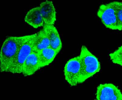
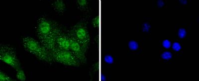
Hela cell; 4% Paraformaldehyde-fixed; Triton X-100 at room temperature for 20 min; Blocking buffer (normal goat serum, SLC0005) at 37°C for 20 min; Antibody incubation with (Aurora A) monoclonal Antibody, Unconjugated (SLM52018R) 1:50, 90 minutes at 37°C; followed by a conjugated Goat Anti-Rabbit IgG antibody at 37°C for 90 minutes, DAPI (blue, C02-04002) was used to stain the cell nuclei.
NIH/3T3 cell; 4% Paraformaldehyde-fixed; Triton X-100 at room temperature for 20 min; Blocking buffer (normal goat serum, SLC0005) at 37°C for 20 min; Antibody incubation with (Aurora A) monoclonal Antibody, Unconjugated (SLM52018R) 1:50, 90 minutes at 37°C; followed by a conjugated Goat Anti-Rabbit IgG antibody at 37°C for 90 minutes, DAPI (blue, C02-04002) was used to stain the cell nuclei.
SKOSLV3 cell; 4% Paraformaldehyde-fixed; Triton X-100 at room temperature for 20 min; Blocking buffer (normal goat serum, SLC0005) at 37°C for 20 min; Antibody incubation with (Aurora A) monoclonal Antibody, Unconjugated (SLM52018R) 1:50, 90 minutes at 37°C; followed by a conjugated Goat Anti-Rabbit IgG antibody at 37°C for 90 minutes, DAPI (blue, C02-04002) was used to stain the cell nuclei.
Blank control: Hela.
Primary Antibody (green line): Rabbit Anti-Aurora A antibody (SLM52018R)
Dilution: 1:50;
Isotype Control Antibody (orange line): Rabbit IgG .
Secondary Antibody : Goat anti-rabbit IgG-AF488
Dilution: 1:1000.
Protocol
The cells were fixed with 4% PFA (10min at room temperature)and then permeabilized with 0.1% PBST for 20 min at room temperature. The cells were then incubated in 5%BSA to block non-specific protein-protein interactions for 30 min at room temperature .Cells stained with Primary Antibody for 30 min at room temperature. The secondary antibody used for 40 min at room temperature. Acquisition of 20,000 events was performed.
Product Feedback Wall
Message :
Your Email :
Copyright © 2007-2018 Sunlong Medical All Rights Reserved.