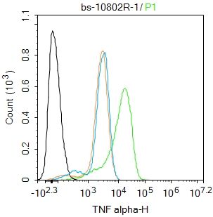
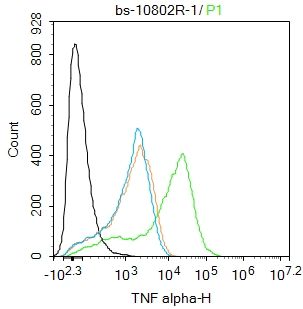
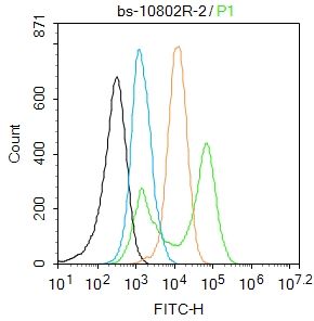
[IF=5.893] WB, IHC ; mouse, human.
[IF=14.593] Bin Wang. et al. Targeted delivery of a STING agonist to brain tumors using bioengineered protein nanoparticles for enhanced immunotherapy. Bioact Mater. 2022 Mar;: IF ; Mouse.
[IF=7.813] Shengyan Yang. et al. Extracellular vesicles delivering nuclear factor I/C for hard tissue engineering: Treatment of apical periodontitis and dentin regeneration:. J Tissue Eng. 2022;(): IHC ; Rat.
[IF=6.529] Mennatallah A. Gowayed. et al. The α7 nAChR allosteric modulator PNU-120596 amends neuroinflammatory and motor consequences of parkinsonism in rats: Role of JAK2/NF-κB/GSk3β/ TNF-α pathway. Biomed Pharmacother. 2022 Apr;148:112776 IHC ; Rat.
[IF=5.59] Li, Jiayi. et al. Activation of the GPX4/TLR4 Signaling Pathway Participates in the Alleviation of Selenium Yeast on Deltamethrin-Provoked Cerebrum Injury in Quails. Mol Neurobiol. 2022 Mar;:1-16 WB ; Quails.
[IF=4.36] Hong Zhao. et al. Protective effect of flavonoids extract of Hippophae rhamnoides L. on alcoholic fatty liver disease through regulating intestinal flora and inhibiting TAK1/p38MAPK/p65NF-κB pathway. J Ethnopharmacol. 2022 Jun;292:115225 WB ; Mouse.
[IF=2.408] Jingru Qin. et al. In-silico analysis identifies the anti-liver injury targets of diammonium glycyrrhizinate: validated in perfluorooctanoic acid-lesioned mouse model. 2022 Mar 31 IHC ; Mouse.
[IF=2.248] Wang Yulong. et al. LncRNA GAS5 promotes abdominal aortic aneurysm formation through regulating the miR-185-5p/ADCY7 axis. Anti-Cancer Drug. 2022 Mar;33(3):225-234 WB ; Human.
[IF=3.905] Fen Xu. et al. Lipoxin A4 and its analog attenuate high fat diet-induced atherosclerosis via Keap1/Nrf2 pathway. Exp Cell Res. 2022 Mar;412:113025 IF ; Rat.
[IF=9.229] Jiarong Yang. et al. Mitigation Effects of Selenium Nanoparticles on Depression-Like Behavior Induced by Fluoride in Mice via the JAK2-STAT3 Pathway. Acs Appl Mater Inter. 2022;14(3):3685–3700 WB ; Mouse.
[IF=7.658] Yongbo Kang. et al. Lactobacillus acidophilus ameliorates obesity in mice through modulation of gut microbiota dysbiosis and intestinal permeability. Pharmacol Res. 2021 Dec;:106020 IHC ; Mouse.
[IF=3.24] Chang Chen. et al. H2O2 gel bleaching induces cytotoxicity and pain conduction in dental pulp stem cells via intracellular reactive oxygen species on enamel/dentin disc. Plos One. 2021 Sep;16(9):e0257221 IF ; rat.
[IF=6.291] Si-Cheng Zhao. et al. Nickel sulfate exposure induces ovarian inflammation and fibrosis and decreases oocyte quality in mice. Ecotox Environ Safe. 2021 Nov;224:112634 IHC ; Mouse.
[IF=1.813] Jiang Yan. et al. Exploring the Mechanism of Berberine Intervention in Ulcerative Colitis from the Perspective of Inflammation and Immunity Based on Systemic Pharmacology. Evid-Based Compl Alt. 2021;2021:997048 IHC ; Rat.
[IF=3.216] Ji-sheng Wang. et al. Study on the Mechanism of Jiawei Shengjiang Powder in Improving Male Asthma-Induced Asthenospermia Based on Network Pharmacology and Bioinformatics. Drug Des Dev Ther. 2021 Mar;15:1245-1259 WB ; Rat.
[IF=5.893] Ju-Tao Yu. et al. DNA methylation of FTO promotes renal inflammation by enhancing m6A of PPAR-α in alcohol-induced kidney injury. Pharmacol Res. 2021 Jan;163:105286 IHC ; Mouse.
[IF=1.918] Wang J et al. Effect of Asthma on Erectile Dysfunction in Rats as Determined by Biological Network AnalysisMed Sci Monit.2020 Dec 20;26:e927491. WB ; Rat.
[IF=1.574] Song L et al. Schisandrin ameliorates cognitive deficits, endoplasmic reticulum stress and neuroinflammation in STZ-induced Alzheimer's disease rats. Exp Anim
. 2020 Aug 5;69(3):363-373. IHC ; rat.
[IF=2.998] Lee M et al. Acute changes in the retina and central retinal artery with methamphetamine. Exp Eye Res. 2020 Feb 7;193:107964. WB&IHF-P ; Mouse.
[IF=0] Artono Surarto B et al. The Association of IL-1 Alpha Level and TNF Alpha Expressions on Bone Destruction in Chronic Suppurative Otitis Media and Cholesteatoma. Indian Journal of Otolaryngology and Head & Neck Surgery.2019. IHC ; Human.
[IF=3.265] Meng HW et al. 4-Methylcoumarin-[5,6-g]-hesperetin attenuates inflammatory responses in alcoholic hepatitis through PPAR-γ activation. Toxicology. 2019 Apr 3;421:9-21. WB ; Mouse.
[IF=7.233] Chen X et al.
A photothermal-triggered nitric oxide nanogenerator combined with siRNA for precise therapy of osteoarthritis by suppressing macrophage inflammation.Nanoscale. 2019 Apr 4;11(14):6693-6709. IHSLCP&IF ; Mouse.
[IF=3.83] Cheng et al. miR-203 Inhibits Alcohol-Induced Hepatic Steatosis by Targeting Lipin1. (2018) Front.Pharmacol. 9:275 WB ; Mouse.


