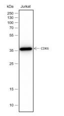
Blocking buffer: 5% NFDM/TBST
Primary Ab dilution: 1:2000
Primary Ab incubation condition: 2 hours at
room temperature
Secondary Ab: Goat Anti-Rabbit IgG H&L
(HRP)
Lysate: Jurkat
Protein loading quantity: 20 μg
Exposure time: 60 s
Predicted MW: 37 kDa
Observed MW: 37 kDa
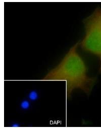
Cell line: HeLa
Fixative: 100% Ice-cold methanol
Permeabilization: 0.1% TritonX-100
Primary Ab dilution: 1:50
Primary incubation condition: 4°C overnight
Secondary Ab: Goat Anti-Rabbit IgG
Nuclear counter stain: DAPI (Blue)
Counter stain: Tubulin (Red)
Comment: Color green is the positive signal for SLM52030R
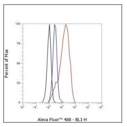
Cell line: HeLa
Fixation: 4% Paraformaldehyde
Permeabilization: 90% Methanol
Primary Ab dilution: 1:50
Secondary Ab: Goat Anti-Rabbit IgG
Unlabelled control: The cell without incubation
with primary antibody and secondary antibody
(Black line).
Isotype control: Rabbit monoclonal IgG (Blue
line).
Comment: Line red is the positive signal for SLM52030R
|