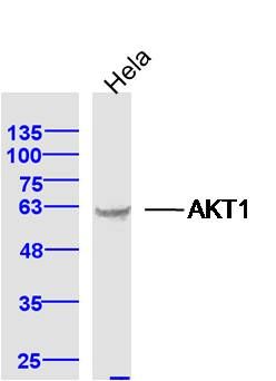
Sample: Hela Cell (Human) Lysate at 40 ug
Primary: Anti-AKT1 (SL0115M) at 1/300 dilution
Secondary: IRDye800CW Goat Anti-Mouse IgG at 1/20000 dilution
Predicted band size: 56 kD
Observed band size: 60 kD
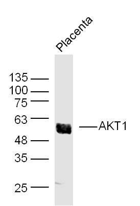
Sample: Placenta (Mouse) Lysate at 30 ug
Primary: Anti- AKT1 (SLM0115M) at 1/300 dilution
Secondary: IRDye800CW Goat Anti-Mouse IgG at 1/20000 dilution
Predicted band size: 56 kD
Observed band size: 56 kD
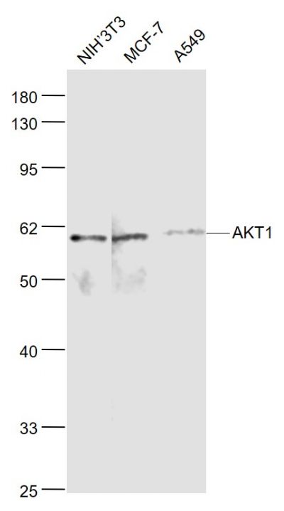
Sample:
NIH/3T3(Mouse) Cell Lysate at 30 ug
MCF-7(Human) Cell Lysate at 30 ug
A549(Human) Cell Lysate at 30 ug
Primary: Anti-AKT1 (SL0115M) at 1/1000 dilution
Secondary: IRDye800CW Goat Anti-Mouse IgG at 1/20000 dilution
Predicted band size: 60 kD
Observed band size: 60 kD
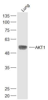
Sample:
Lung (Mouse) Lysate at 40 ug
Primary: Anti-AKT1 (SL0115M) at 1/300 dilution
Secondary: IRDye800CW Goat Anti-Rabbit IgG at 1/20000 dilution
Predicted band size: 56 kD
Observed band size: 56 kD
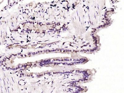
Paraformaldehyde-fixed, paraffin embedded (rat lung); Antigen retrieval by boiling in sodium citrate buffer (pH6.0) for 15min; Block endogenous peroxidase by 3% hydrogen peroxide for 20 minutes; Blocking buffer (normal goat serum) at 37°C for 30min; Antibody incubation with (AKT1) Monoclonal Antibody, Unconjugated (SL0115M) at 1:200 overnight at 4°C, followed by operating according to SP Kit(Mouse)(sp-0024) instructionsand DAB staining.
Paraformaldehyde-fixed, paraffin embedded (mouse liver); Antigen retrieval by boiling in sodium citrate buffer (pH6.0) for 15min; Block endogenous peroxidase by 3% hydrogen peroxide for 20 minutes; Blocking buffer (normal goat serum) at 37°C for 30min; Antibody incubation with (AKT1) Polyclonal Antibody, Unconjugated (SL0115M) at 1:200 overnight at 4°C, followed by operating according to SP Kit(Rabbit) (sp-0024) instructionsand DAB staining.
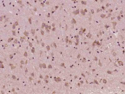
Paraformaldehyde-fixed, paraffin embedded (Mouse brain); Antigen retrieval by boiling in sodium citrate buffer (pH6.0) for 15min; Block endogenous peroxidase by 3% hydrogen peroxide for 20 minutes; Blocking buffer (normal goat serum) at 37°C for 30min; Antibody incubation with (AKT1) Monoclonal Antibody, Unconjugated (SL0115M) at 1:400 overnight at 4°C, followed by operating according to SP Kit(Mouse) (sp-0024) instructionsand DAB staining.
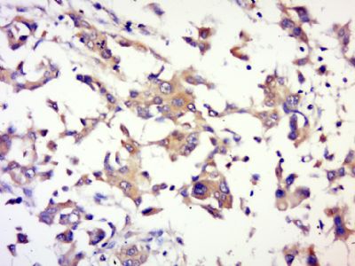
Paraformaldehyde-fixed, paraffin embedded (human memmery cancer); Antigen retrieval by boiling in sodium citrate buffer (pH6.0) for 15min; Block endogenous peroxidase by 3% hydrogen peroxide for 20 minutes; Blocking buffer (normal goat serum) at 37°C for 30min; Antibody incubation with (AKT1) Polyclonal Antibody, Unconjugated (SL0115R) at 1:500 overnight at 4°C, followed by a conjugated secondary (sp-0023) for 20 minutes and DAB staining.
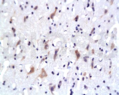
Tissue/cell: rat brain tissue; 4% Paraformaldehyde-fixed and paraffin-embedded;
Antigen retrieval: citrate buffer ( 0.01M, pH 6.0 ), Boiling bathing for 15min; Block endogenous peroxidase by 3% Hydrogen peroxide for 30min; Blocking buffer (normal goat serum,SLC0005) at 37∩ for 20 min;
Incubation: Anti-PKB Polyclonal Antibody, Unconjugated(SL0115M) 1:200, overnight at 4∑C, followed by conjugation to the secondary antibody(SP-0024) and DAB(SLC0010) staining
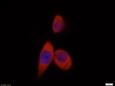
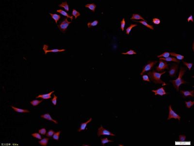
Tissue/cell:MCF7 cell; 4% Paraformaldehyde-fixed; Triton X-100 at room temperature for 20 min; Blocking buffer (normal goat serum, SLC0005) at 37°C for 20 min; Antibody incubation with (AKT1) polyclonal Antibody, Unconjugated (SL0115M) 1:100, 90 minutes at 37°C; followed by a CY3 conjugated Goat Anti-Mouse IgG antibody at 37°C for 90 minutes, DAPI (blue, C02-04002) was used to stain the cell nuclei.
Tissue/cell:MCF7 cell; 4% Paraformaldehyde-fixed; Triton X-100 at room temperature for 20 min; Blocking buffer (normal goat serum, SLC0005) at 37°C for 20 min; Antibody incubation with (AKT1) polyclonal Antibody, Unconjugated (SL0115M) 1:100, 90 minutes at 37°C; followed by a CY3 conjugated Goat Anti-Mouse IgG antibody at 37°C for 90 minutes, DAPI (blue, C02-04002) was used to stain the cell nuclei.
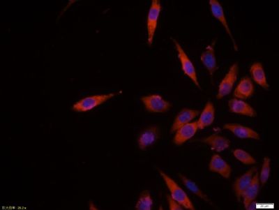
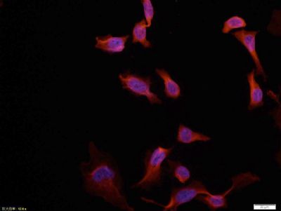
Tissue/cell: Hela cell; 4% Paraformaldehyde-fixed; Triton X-100 at room temperature for 20 min; Blocking buffer (normal goat serum, SLC0005) at 37°C for 20 min; Antibody incubation with (MAKT1) polyclonal Antibody, Unconjugated (SL0115M) 1:100, 90 minutes at 37°C; followed by a conjugated Goat Anti-Mouse IgG-CY3 antibody at 37°C for 90 minutes, DAPI (blue, C02-04002) was used to stain the cell nuclei.
Tissue/cell: Hela cell; 4% Paraformaldehyde-fixed; Triton X-100 at room temperature for 20 min; Blocking buffer (normal goat serum, SLC0005) at 37°C for 20 min; Antibody incubation with (MAKT1) polyclonal Antibody, Unconjugated (SL0115M) 1:100, 90 minutes at 37°C; followed by a conjugated Goat Anti-Mouse IgG-CY3 antibody at 37°C for 90 minutes, DAPI (blue, C02-04002) was used to stain the cell nuclei.
|