Home > Product > Antibody > Mouse Anti-Cystatin C antibody
Cystatin-3; Cystatin3; Cystatin 3; CystatinC; CST 3; CST3; CST-3; Cystatin C; AD 8; AD8; Amyloid angiopathy and cerebral hemorrhage; Cst 3; Cst3; CST3 protein; Gamma trace; HCCAA; Neuroendocrine basic polypeptide; Post gamma globulin; ARMD11; MGC117328; C
Cat:
SLM33286M
Species Reactivity:
Human,Mouse,
Immunogen:
Recombinant human Cystatin C Protein
Format:
Liquid
Storage instructions:
Shipped at 4℃. Store at -20 °C for one year. Avoid repeated freeze/thaw cycles.
Concentration:
1mg/ml
Clonality:
Monoclonal
Isotype:
IgG
Applications:
WB=1:500-1000ELISA=1:5000-10000IHC-P=1:500-1000IHC-F=1:500-1000ICC=1:100-500IF=1:500-1000(Paraffin sections need to do antigen repair)not yet tested in other applications.optimal dilutions/concentrations should be determined by the end user.
Host:
Mouse
Product Overview:
Sample: Hela(Human) Cell Lysate at 30 ugHcclm3(Human) Cell Lysate at 30 ugPrimary: Anti-Cystatin C (SLM33286M) at 1/1000 dilutionSecondary: IRDye800CW Goat Anti-Mouse IgG at 1/20000 dilutionPredicted band size: 14 kDObserved band size: 14 kDSample: 293T(Human) Cell Lysate at 30 ugPrimary: Anti-Cystatin C (SLM33286M) at 1/1000 dilutionSecondary: IRDye800CW Goat Anti-Mouse IgG at 1/20000 dilutionPredicted band size: 14 kDObserved band size: 14 kDSample: A549(Human) Cell Lysate at 30 ugU937(Human) Cell Lysate at 30 ugPrimary: Anti- Cystatin C (SLM33286M) at 1/1000 dilutionSecondary: IRDye800CW Goat Anti-Mouse IgG at 1/20000 dilutionPredicted band size: 14 kDObserved band size: 15 kDParaformaldehyde-fixed, paraffin embedded (Human kidney tissue); Antigen retrieval by boiling in sodium citrate buffer (pH6.0) for 15min; Block endogenous peroxidase by 3% hydrogen peroxide for 20 minutes; Blocking buffer (normal goat serum) at 37°C for 30min; Antibody incubation with (Cystatin C) Monoclonal Antibody, Unconjugated (SLM33286M) at 1:400 overnight at 4°C, followed by operating according to SP Kit(Mouse)(sp-0024) instructionsand DAB staining.Paraformaldehyde-fixed, paraffin embedded (human brain glioma); Antigen retrieval by boiling in sodium citrate buffer (pH6.0) for 15min; Block endogenous peroxidase by 3% hydrogen peroxide for 20 minutes; Blocking buffer (normal goat serum) at 37°C for 30min; Antibody incubation with (Cystatin C) Monoclonal Antibody, Unconjugated (SLM33286M) at 1:400 overnight at 4°C, followed by operating according to SP Kit(Mouse)(sp-0024) instructionsand DAB staining.
More
Unit:
Price: $
Product PDFs
Datasheet:


The cystatin superfamily encompasses proteins that contain multiple cystatin-like sequences. Some of the members are active cysteine protease inhibitors, while others have lost or perhaps never acquired this inhibitory activity. There are three inhibitory families in the superfamily, including the type 1 cystatins(stefins), type 2 cystatins and the kininogens. The type 2 cystatin proteins are a class of cysteine proteinase inhibitors found in a variety of human fluids and secretions, where they appear to provide protective functions. The cystatin locus on chromosome 20 contains the majority of the type 2 cystatin genes and pseudogenes. This gene is located in the cystatin locus and encodes the most abundant extracellular inhibitor of cysteine proteases, which is found in high concentrations in biological fluids and is expressed in virtually all organs of the body. A mutation in this gene has been associated with amyloid angiopathy. Expression of this protein in vascular wall smooth muscle cells is severely reduced in both atherosclerotic and aneurysmal aortic lesions, establishing its role in vascular disease. [provided by RefSeq].

Function:
As an inhibitor of cysteine proteinases, this protein is thought to serve an important physiological role as a local regulator of this enzyme activity.

Subunit:
Expressed in submandibular and sublingual saliva but not in parotid saliva (at protein level). Expressed in various body fluids, such as the cerebrospinal fluid and plasma. Expressed in highest levels in the epididymis, vas deferens, brain, thymus, and ovary and the lowest in the submandibular gland.

Subcellular Location:
Secreted.

DISEASE:
Defects in CST3 are the cause of amyloidosis type 6 (AMYL6) [MIM:105150]; also known as hereditary cerebral hemorrhage with amyloidosis (HCHWA), cerebral amyloid angiopathy (CAA) or cerebroarterial amyloidosis Icelandic type. AMYL6 is a hereditary generalized amyloidosis due to cystatin C amyloid deposition. Cystatin C amyloid accumulates in the walls of arteries, arterioles, and sometimes capillaries and veins of the brain, and in various organs including lymphoid tissue, spleen, salivary glands, and seminal vesicles. Amyloid deposition in the cerebral vessels results in cerebral amyloid angiopathy, cerebral hemorrhage and premature stroke. Cystatin C levels in the cerebrospinal fluid are abnormally low.

Similarity:
Belongs to the cystatin family.

SWISS:
P01034

Gene ID:
1471

Database links:

Entrez Gene: 1471 Human

Entrez Gene: 13010 Mouse

Entrez Gene: 25307 Rat

Omim: 604312 Human

SwissProt: P01034 Human

SwissProt: P2192 Mouse

SwissProt: P14841 Rat

Unigene: 304682 Human

Unigene: 4263 Mouse

Unigene: 106351 Rat


胱抑素C( cystatin c)是一种半胱氨酸蛋白酶抑制剂,也被称为γ-微量蛋白及γ-后球蛋白,广泛存在于各种组织的有核细胞和体液中,是一种低分子量、碱性非糖化蛋白质,分子量为13.3KD,由122个氨基酸残基组成,可由机体所有有核细胞产生,产生率恒定。循环中的胱抑素c仅经肾小球滤过而被清除,是一种反映肾小球滤过率变化的内源性标志物,并在近曲小管重吸收,但重吸收后被完全代谢分解,不返回血液,因此,其血中浓度由肾小球滤过决定,而不依赖任何外来因素,如性别、年龄、饮食的影响,是一种反映肾小球滤过率变化的理想同源性标志物。 Picture

Sample:
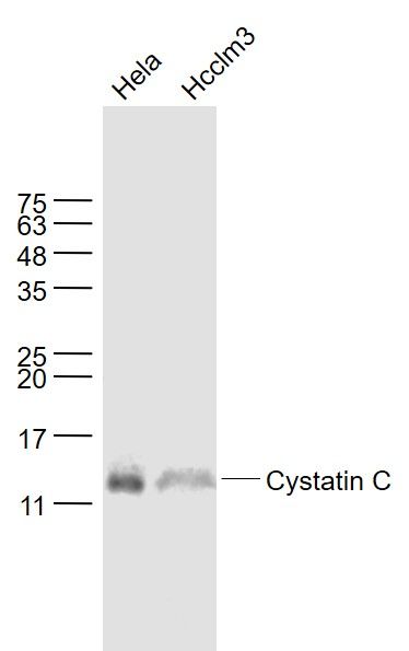
Hela(Human) Cell Lysate at 30 ug
Hcclm3(Human) Cell Lysate at 30 ug
Primary: Anti-Cystatin C (SLM33286M) at 1/1000 dilution
Secondary: IRDye800CW Goat Anti-Mouse IgG at 1/20000 dilution
Predicted band size: 14 kD
Observed band size: 14 kD
Sample:
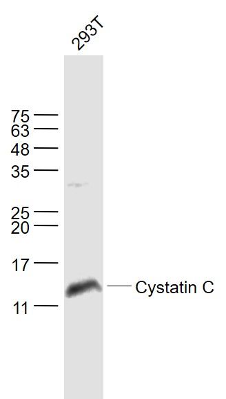
293T(Human) Cell Lysate at 30 ug
Primary: Anti-Cystatin C (SLM33286M) at 1/1000 dilution
Secondary: IRDye800CW Goat Anti-Mouse IgG at 1/20000 dilution
Predicted band size: 14 kD
Observed band size: 14 kD
Sample:
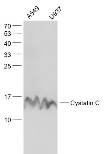
A549(Human) Cell Lysate at 30 ug
U937(Human) Cell Lysate at 30 ug
Primary: Anti- Cystatin C (SLM33286M) at 1/1000 dilution
Secondary: IRDye800CW Goat Anti-Mouse IgG at 1/20000 dilution
Predicted band size: 14 kD
Observed band size: 15 kD
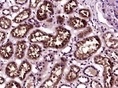
Paraformaldehyde-fixed, paraffin embedded (Human kidney tissue); Antigen retrieval by boiling in sodium citrate buffer (pH6.0) for 15min; Block endogenous peroxidase by 3% hydrogen peroxide for 20 minutes; Blocking buffer (normal goat serum) at 37°C for 30min; Antibody incubation with (Cystatin C) Monoclonal Antibody, Unconjugated (SLM33286M) at 1:400 overnight at 4°C, followed by operating according to SP Kit(Mouse)(sp-0024) instructionsand DAB staining.
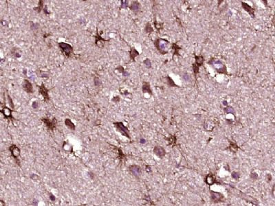
Paraformaldehyde-fixed, paraffin embedded (human brain glioma); Antigen retrieval by boiling in sodium citrate buffer (pH6.0) for 15min; Block endogenous peroxidase by 3% hydrogen peroxide for 20 minutes; Blocking buffer (normal goat serum) at 37°C for 30min; Antibody incubation with (Cystatin C) Monoclonal Antibody, Unconjugated (SLM33286M) at 1:400 overnight at 4°C, followed by operating according to SP Kit(Mouse)(sp-0024) instructionsand DAB staining.
Product Feedback Wall
Message :
Your Email :
Copyright © 2007-2018 Sunlong Medical All Rights Reserved.