Home > Product > Antibody > Mouse Anti-Cytochrome C antibody
CytC; CYC; CYCS; Cytochrome c somatic; HCS; CYC_HUMAN; Cytochrome c; MSA06; THC4.
Cat:
SLM33193M
Species Reactivity:
Human,Rat,(predicted: Mouse,)
Immunogen:
Recombinant human Cytochrome C Protein
Format:
Liquid
Storage instructions:
Shipped at 4℃. Store at -20 °C for one year. Avoid repeated freeze/thaw cycles.
Concentration:
1mg/ml
Clonality:
Monoclonal
Isotype:
IgG
Applications:
WB=1:500-1000IHC-P=1:100-500IHC-F=1:100-500ICC=1:100IF=1:100-500(Paraffin sections need to do antigen repair)not yet tested in other applications.optimal dilutions/concentrations should be determined by the end user.
Host:
Mouse
Product Overview:
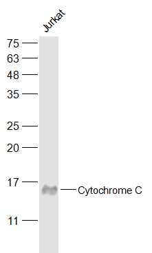
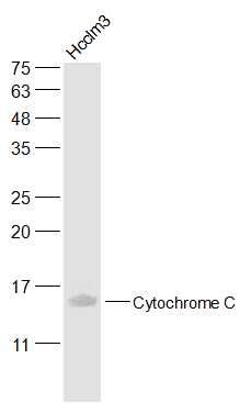
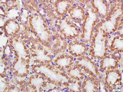
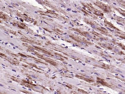
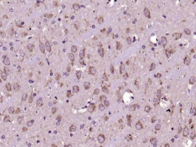
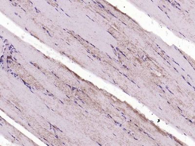
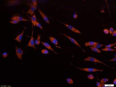
Sample: Jurkat(Human) Cell Lysate at 30 ugPrimary: Anti-Cytochrome C (SLM33193M) at 1/50000 dilutionSecondary: IRDye800CW Goat Anti-Mouse IgG at 1/20000 dilutionPredicted band size: 14 kDObserved band size: 14 kDSample: Hcclm3(Human) Cell Lysate at 30 ugPrimary: Anti-Cytochrome C (SLM33193M) at 1/100000 dilutionSecondary: IRDye800CW Goat Anti-Mouse IgG at 1/20000 dilutionPredicted band size: 14 kDObserved band size: 14 kDParaformaldehyde-fixed, paraffin embedded (Rat kidney); Antigen retrieval by boiling in sodium citrate buffer (pH6.0) for 15min; Block endogenous peroxidase by 3% hydrogen peroxide for 20 minutes; Blocking buffer (normal goat serum) at 37°C for 30min; Antibody incubation with (Cytochrome C) Monoclonal Antibody, Unconjugated (SLM33193M) at 1:400 overnight at 4°C, followed by a conjugated secondary (sp-0023) for 20 minutes and DAB staining.Paraformaldehyde-fixed, paraffin embedded (rat heart); Antigen retrieval by boiling in sodium citrate buffer (pH6.0) for 15min; Block endogenous peroxidase by 3% hydrogen peroxide for 20 minutes; Blocking buffer (normal goat serum) at 37°C for 30min; Antibody incubation with (Cytochrome C) Monoclonal Antibody, Unconjugated (SLM33193M) at 1:200 overnight at 4°C, followed by operating according to SP Kit(Mouse)(sp-0024) instructionsand DAB staining.Paraformaldehyde-fixed, paraffin embedded (rat brain); Antigen retrieval by boiling in sodium citrate buffer (pH6.0) for 15min; Block endogenous peroxidase by 3% hydrogen peroxide for 20 minutes; Blocking buffer (normal goat serum) at 37°C for 30min; Antibody incubation with (Cytochrome C) Monoclonal Antibody, Unconjugated (SLM33193M) at 1:200 overnight at 4°C, followed by operating according to SP Kit(Mouse)(sp-0024) instructionsand DAB staining.Paraformaldehyde-fixed, paraffin embedded (rat skeletal muscle); Antigen retrieval by boiling in sodium citrate buffer (pH6.0) for 15min; Block endogenous peroxidase by 3% hydrogen peroxide for 20 minutes; Blocking buffer (normal goat serum) at 37°C for 30min; Antibody incubation with (Cytochrome C) Monoclonal Antibody, Unconjugated (SLM33193M) at 1:200 overnight at 4°C, followed by operating according to SP Kit(Mouse)(sp-0024) instructionsand DAB staining.SH-SY5Y cell; 4% Paraformaldehyde-fixed; Triton X-100 at room temperature for 20 min; Blocking buffer (normal goat serum, SLC0005) at 37°C for 20 min; Antibody incubation with (Cytochrome C) monoclonal Antibody, Unconjugated (SLM33193M) 1:100, 90 minutes at 37°C; followed by a conjugated Goat Anti-Mouse IgG antibody at 37°C for 90 minutes, DAPI (blue, C02-04002) was used to stain the cell nuclei.
More
Unit:
Price: $
Product PDFs
Datasheet:


Cytochrome C is an electron transporting protein that resides within the intermembrane space of the mitochondria, where it plays a critical role in the process of oxidative phosphorylation and production of cellular ATP. An increasing amount of interest has been directed toward the role which cytocrome C has been demonstrated to play in apoptotic processes. Following exposure to apoptotic stimuli, cytochrome C is rapidly released from the mitochondria into the cytosol, an event which may be required for the completion of apoptosis in some systems. Cytosolic cytochrome C functions in the activation of caspase 3, an ICE family molecule that is a key effector of apoptosis.

Function:
Electron carrier protein. The oxidized form of the cytochrome c heme group can accept an electron from the heme group of the cytochrome c1 subunit of cytochrome reductase. Cytochrome c then transfers this electron to the cytochrome oxidase complex, the final protein carrier in the mitochondrial electron-transport chain.
Plays a role in apoptosis. Suppression of the anti-apoptotic members or activation of the pro-apoptotic members of the Bcl-2 family leads to altered mitochondrial membrane permeability resulting in release of cytochrome c into the cytosol. Binding of cytochrome c to Apaf-1 triggers the activation of caspase-9, which then accelerates apoptosis by activating other caspases.

Subcellular Location:
Mitochondrion intermembrane space. Note=Loosely associated with the inner membrane.

Post-translational modifications:
Binds 1 heme group per subunit.
Phosphorylation at Tyr-49 and Tyr-98 both reduce by half the turnover in the reaction with cytochrome c oxidase, down-regulating mitochondrial respiration.

DISEASE:
Defects in CYCS are the cause of thrombocytopenia type 4 (THC4) [MIM:62404]; also known as autosomal dominant thrombocytopenia type 4. Thrombocytopenia is the presence of relatively few platelets in blood. THC4 is a non-syndromic form of thrombocytopenia. Clinical manifestations of thrombocytopenia are absent or mild. THC4 may be caused by dysregulated platelet formation.

Similarity:
Belongs to the cytochrome c family.

SWISS:
P99999

Gene ID:
54205

Database links:

Entrez Gene: 54205 Human

Entrez Gene: 13063 Mouse

Entrez Gene: 25309 Rat

Omim: 123970 Human

SwissProt: P99999 Human

SwissProt: P62897 Mouse

SwissProt: P62898 Rat

Unigene: 437060 Human


细胞色素C(cytC)是一种电子传递链蛋白为线粒体呼吸链必须的成份之一。在哺乳动物细胞中,如此高度保守性蛋白常分布在线粒体内膜。
新近研究证明细胞浆中细胞色素C为激活细胞调亡所必需的因子。在调亡的过程中,细胞色素C从线粒体膜被易位到细胞浆,由细胞色素C激活Caspase-3(CPP32)。
细胞色素C的易位可被过量表达的Bcl-2阻断。细胞色素B与细胞色素C1和Rieske蛋白相结合而形成复合物III(也称细胞色素B-C1复合物)参与细胞呼吸链。该蛋白动物种属间同源性较高;如 :猪、犬、牛、鸡、豚鼠等。
Picture

Sample:
Jurkat(Human) Cell Lysate at 30 ug
Primary: Anti-Cytochrome C (SLM33193M) at 1/50000 dilution
Secondary: IRDye800CW Goat Anti-Mouse IgG at 1/20000 dilution
Predicted band size: 14 kD
Observed band size: 14 kD
Sample:
Hcclm3(Human) Cell Lysate at 30 ug
Primary: Anti-Cytochrome C (SLM33193M) at 1/100000 dilution
Secondary: IRDye800CW Goat Anti-Mouse IgG at 1/20000 dilution
Predicted band size: 14 kD
Observed band size: 14 kD
Paraformaldehyde-fixed, paraffin embedded (Rat kidney); Antigen retrieval by boiling in sodium citrate buffer (pH6.0) for 15min; Block endogenous peroxidase by 3% hydrogen peroxide for 20 minutes; Blocking buffer (normal goat serum) at 37°C for 30min; Antibody incubation with (Cytochrome C) Monoclonal Antibody, Unconjugated (SLM33193M) at 1:400 overnight at 4°C, followed by a conjugated secondary (sp-0023) for 20 minutes and DAB staining.
Paraformaldehyde-fixed, paraffin embedded (rat heart); Antigen retrieval by boiling in sodium citrate buffer (pH6.0) for 15min; Block endogenous peroxidase by 3% hydrogen peroxide for 20 minutes; Blocking buffer (normal goat serum) at 37°C for 30min; Antibody incubation with (Cytochrome C) Monoclonal Antibody, Unconjugated (SLM33193M) at 1:200 overnight at 4°C, followed by operating according to SP Kit(Mouse)(sp-0024) instructionsand DAB staining.
Paraformaldehyde-fixed, paraffin embedded (rat brain); Antigen retrieval by boiling in sodium citrate buffer (pH6.0) for 15min; Block endogenous peroxidase by 3% hydrogen peroxide for 20 minutes; Blocking buffer (normal goat serum) at 37°C for 30min; Antibody incubation with (Cytochrome C) Monoclonal Antibody, Unconjugated (SLM33193M) at 1:200 overnight at 4°C, followed by operating according to SP Kit(Mouse)(sp-0024) instructionsand DAB staining.
Paraformaldehyde-fixed, paraffin embedded (rat skeletal muscle); Antigen retrieval by boiling in sodium citrate buffer (pH6.0) for 15min; Block endogenous peroxidase by 3% hydrogen peroxide for 20 minutes; Blocking buffer (normal goat serum) at 37°C for 30min; Antibody incubation with (Cytochrome C) Monoclonal Antibody, Unconjugated (SLM33193M) at 1:200 overnight at 4°C, followed by operating according to SP Kit(Mouse)(sp-0024) instructionsand DAB staining.
SH-SY5Y cell; 4% Paraformaldehyde-fixed; Triton X-100 at room temperature for 20 min; Blocking buffer (normal goat serum, SLC0005) at 37°C for 20 min; Antibody incubation with (Cytochrome C) monoclonal Antibody, Unconjugated (SLM33193M) 1:100, 90 minutes at 37°C; followed by a conjugated Goat Anti-Mouse IgG antibody at 37°C for 90 minutes, DAPI (blue, C02-04002) was used to stain the cell nuclei.
Product Feedback Wall
Message :
Your Email :
Copyright © 2007-2018 Sunlong Medical All Rights Reserved.