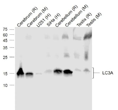
Sample:
Lane 1: Cerebrum (Rat) Lysate at 40 ug
Lane 2: Cerebrum (Mouse) Lysate at 40 ug
Lane 3: U251 (Human) Cell Lysate at 30 ug
Lane 4: SiHa (Human) Cell Lysate at 30 ug
Lane 5: Cerebellum (Rat) Lysate at 40 ug
Lane 6: Cerebellum (Mouse) Lysate at 40 ug
Lane 7: Testis (Rat) Lysate at 40 ug
Lane 8: Testis (Mouse) Lysate at 40 ug
Primary: Anti-LC3A (SLM33309M) at 1/1000 dilution
Secondary: IRDye800CW Goat Anti-Mouse IgG at 1/20000 dilution
Predicted band size: 17 kD
Observed band size: 15 kD
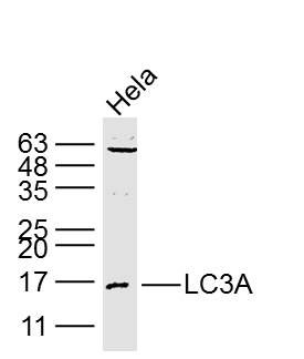
Sample:
Hela Cell (Human) Lysate at 40 ug
Primary: Anti- LC3A (SLM33309M) at 1/1000 dilution
Secondary: IRDye800CW Goat Anti-Mouse IgG at 1/20000 dilution
Predicted band size: 14/16 kD
Observed band size: 16 kD
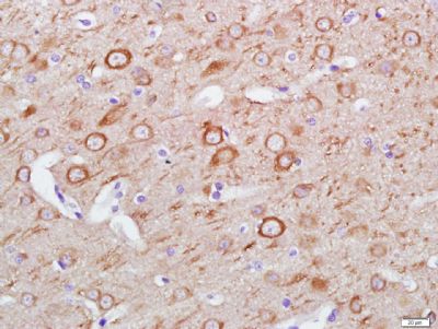
Paraformaldehyde-fixed, paraffin embedded (Rat brain); Antigen retrieval by boiling in sodium citrate buffer (pH6.0) for 15min; Block endogenous peroxidase by 3% hydrogen peroxide for 20 minutes; Blocking buffer (normal goat serum) at 37°C for 30min; Antibody incubation with (LC3A) Monoclonal Antibody, Unconjugated (SLM33309M) at 1:400 overnight at 4°C, followed by a conjugated secondary (sp-0023) for 20 minutes and DAB staining.
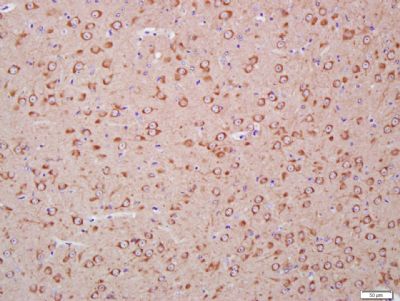
Paraformaldehyde-fixed, paraffin embedded (Mouse brain); Antigen retrieval by boiling in sodium citrate buffer (pH6.0) for 15min; Block endogenous peroxidase by 3% hydrogen peroxide for 20 minutes; Blocking buffer (normal goat serum) at 37°C for 30min; Antibody incubation with (LC3A) Monoclonal Antibody, Unconjugated (SLM33309M) at 1:400 overnight at 4°C, followed by a conjugated secondary (sp-0023) for 20 minutes and DAB staining.
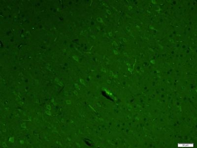
Paraformaldehyde-fixed, paraffin embedded (Rat brain); Antigen retrieval by boiling in sodium citrate buffer (pH6.0) for 15min; Block endogenous peroxidase by 3% hydrogen peroxide for 20 minutes; Blocking buffer (normal goat serum) at 37°C for 30min; Antibody incubation with (LC3A) Monoclonal Antibody, Unconjugated (SLM33309M) at 1:400 overnight at 4°C, followed by a conjugated Goat Anti-Mouse IgG antibody (SL0296G-FITC) for 90 minutes, and DAPI for nuclei staining.
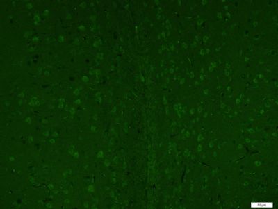
Paraformaldehyde-fixed, paraffin embedded (Mouse brain); Antigen retrieval by boiling in sodium citrate buffer (pH6.0) for 15min; Block endogenous peroxidase by 3% hydrogen peroxide for 20 minutes; Blocking buffer (normal goat serum) at 37°C for 30min; Antibody incubation with (LC3A) Monoclonal Antibody, Unconjugated (SLM33309M) at 1:400 overnight at 4°C, followed by a conjugated Goat Anti-Mouse IgG antibody (SL0296G-FITC) for 90 minutes, and DAPI for nuclei staining.
|