The product encoded by this gene belongs to the actin family of proteins, which are highly conserved proteins that play a role in cell motility, structure and integrity. Alpha, beta and gamma actin isoforms have been identified, with alpha actins being a major constituent of the contractile apparatus, while beta and gamma actins are involved in the regulation of cell motility. This actin is an alpha actin that is found in skeletal muscle. Mutations in this gene cause nemaline myopathy type 3, congenital myopathy with excess of thin myofilaments, congenital myopathy with cores, and congenital myopathy with fiber-type disproportion, diseases that lead to muscle fiber defects. [provided by RefSeq, Jul 2008]
Function:
Actins are highly conserved proteins that are involved in various types of cell motility and are ubiquitously expressed in all eukaryotic cells.
Subunit:
Polymerization of globular actin (G-actin) leads to a structural filament (F-actin) in the form of a two-stranded helix. Each actin can bind to 4 others. Identified in a complex composed of ACTA1, COBL, GSN AND TMSB4X. Interacts with TTID. Interacts (via its SLCterminus) with USP25; the interaction occurs for all USP25 isoforms but is strongest for isoform USP25min muscle differentiating cells.
Subcellular Location:
Cytoplasm, cytoskeleton.
Post-translational modifications:
Oxidation of Met-46 and Met-49 by MICALs (MICAL1, MICAL2 or MICAL3) to form methionine sulfoxide promotes actin filament depolymerization. MICAL1 and MICAL2 produce the (R)-S-oxide form. The (R)-S-oxide form is reverted by MSRB1 and MSRB2, which promote actin repolymerization.
Monomethylation at Lys-86 (K84me1) regulates actin-myosin interaction and actomyosin-dependent processes. Demethylation by ALKSH4 is required for maintaining actomyosin dynamics supporting normal cleavage furrow ingression during cytokinesis and cell migration.
DISEASE:
Nemaline myopathy 3 (NEM3) [MIM:16360]: A form of nemaline myopathy. Nemaline myopathies are muscular disorders characterized by muscle weakness of varying severity and onset, and abnormal thread-like or rod-shaped structures in muscle fibers on histologic examination. Note=The disease is caused by mutations affecting the gene represented in this entry.
Myopathy, actin, congenital, with excess of thin myofilaments (MPCETM) [MIM:16360]: A congenital muscular disorder characterized at histological level by areas of sarcoplasm devoid of normal myofibrils and mitochondria, and replaced with dense masses of thin filaments. Central cores, rods, ragged red fibers and necrosis are absent. Note=The disease is caused by mutations affecting the gene represented in this entry.
Myopathy, congenital, with fiber-type disproportion (CFTD) [MIM:255310]: A genetically heterogeneous disorder in which there is relative hypotrophy of type 1 muscle fibers compared to type 2 fibers on skeletal muscle biopsy. However, these findings are not specific and can be found in many different myopathic and neuropathic conditions. Note=The disease is caused by mutations affecting the gene represented in this entry.
Similarity:
Belongs to the actin family.
SWISS:
P68133
Gene ID:
58
Database links:
Entrez Gene: 421534 Chicken
Entrez Gene: 281592 Cow
Entrez Gene: 58 Human
Entrez Gene: 11459 Mouse
Entrez Gene: 100154254 Pig
Entrez Gene: 29437 Rat
Omim: 102610 Human
SwissProt: P68139 Chicken
SwissProt: P68138 Cow
SwissProt: P68133 Human
SwissProt: P68134 Mouse
SwissProt: P68137 Pig
SwissProt: P68135 Rabbit
SwissProt: P68136 Rat
Unigene: 1288 Human
Unigene: 214950 Mouse
Unigene: 82732 Rat
| Picture |
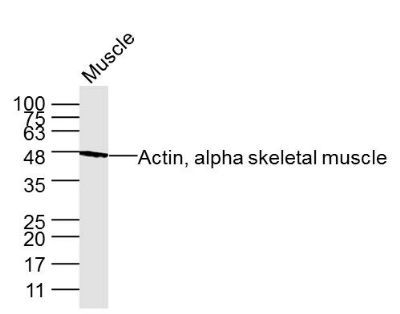
Sample: Muscle (Mouse) Lysate at 40 ug
Primary: Anti- Actin, alpha skeletal muscle (SLM33308M) at 1/2 000 dilution
Secondary: IRDye800CW Goat Anti-Mouse IgG at 1/20000 dilution
Predicted band size: 42 kD
Observed band size: 47 kD
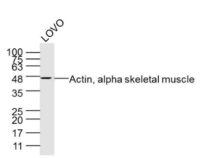
Sample: LOVO Cell (Human) Lysate at 40 ug
Primary: Anti- Actin, alpha skeletal muscle (SLM33308M) at 1/2 000 dilution
Secondary: IRDye800CW Goat Anti-Mouse IgG at 1/20000 dilution
Predicted band size: 42 kD
Observed band size: 47 kD
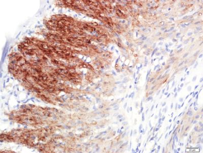
Paraformaldehyde-fixed, paraffin embedded (Rat uterine); Antigen retrieval by boiling in sodium citrate buffer (pH6.0) for 15min; Block endogenous peroxidase by 3% hydrogen peroxide for 20 minutes; Blocking buffer (normal goat serum) at 37°C for 30min; Antibody incubation with (Actin, alpha skeletal muscle) Monoclonal Antibody, Unconjugated (33308M-5E9) at 1:400 overnight at 4°C, followed by a conjugated secondary (sp-0023) for 20 minutes and DAB staining.
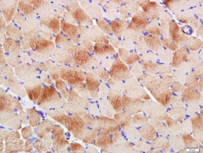
Paraformaldehyde-fixed, paraffin embedded (Rat skeletal muscle); Antigen retrieval by boiling in sodium citrate buffer (pH6.0) for 15min; Block endogenous peroxidase by 3% hydrogen peroxide for 20 minutes; Blocking buffer (normal goat serum) at 37°C for 30min; Antibody incubation with (Actin, alpha skeletal muscle) Monoclonal Antibody, Unconjugated (33308M-5E9) at 1:400 overnight at 4°C, followed by a conjugated secondary (sp-0023) for 20 minutes and DAB staining.
|
|
|