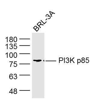
Sample: BRL-3A Cell (Rat) Lysate at 40 ug
Primary: Anti-PI3K p85 (SLM33219M) at 1/1000 dilution
Secondary: IRDye800CW Goat Anti-Mouse IgG at 1/20000 dilution
Predicted band size: 80 kD
Observed band size: 80 kD
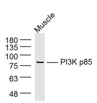
Sample: Muscle (Mouse) Lysate at 40 ug
Primary: Anti-PI3K p85 (SLM33219M) at 1/1000 dilution
Secondary: IRDye800CW Goat Anti-Mouse IgG at 1/20000 dilution
Predicted band size: 80 kD
Observed band size: 80 kD
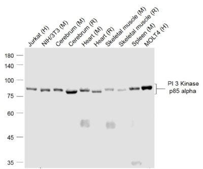
Sample:
Lane 1: Jurkat (Human) Cell Lysate at 30 ug
Lane 2: NIH/3T3(Mouse) Cell Lysate at 30 ug
Lane 3: Cerebrum (Mouse) Lysate at 40 ug
Lane 4: Cerebrum (Rat) Lysate at 40 ug
Lane 5: Heart (Mouse) Lysate at 40 ug
Lane 6: Heart (Rat) Lysate at 40 ug
Lane 7: Skeletal muscle (Mouse) Lysate at 40 ug
Lane 8: Skeletal muscle (Rat) Lysate at 40 ug
Lane 9: Spleen (Mouse) Lysate at 40 ug
Lane 10: MOLT4 (Human) Cell Lysate at 30 ug
Primary: Anti-PI 3 Kinase p85 alpha (SLM33219M) at 1/1000 dilution
Secondary: IRDye800CW Goat Anti-Mouse IgG at 1/20000 dilution
Predicted band size: 85 kD
Observed band size: 85 kD
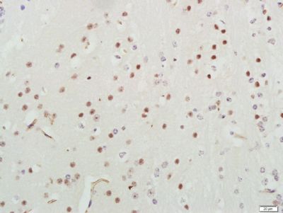
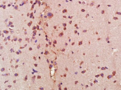
Paraformaldehyde-fixed, paraffin embedded (Mouse brain); Antigen retrieval by boiling in sodium citrate buffer (pH6.0) for 15min; Block endogenous peroxidase by 3% hydrogen peroxide for 20 minutes; Blocking buffer (normal goat serum) at 37°C for 30min; Antibody incubation with (PI3K p85) Monoclonal Antibody, Unconjugated (SLM33219M) at 1:400 overnight at 4°C, followed by a conjugated secondary (sp-0023) for 20 minutes and DAB staining.
Paraformaldehyde-fixed, paraffin embedded (Mouse brain); Antigen retrieval by boiling in sodium citrate buffer (pH6.0) for 15min; Block endogenous peroxidase by 3% hydrogen peroxide for 20 minutes; Blocking buffer (normal goat serum) at 37°C for 30min; Antibody incubation with (PI 3 Kinase p85 alpha) Monoclonal Antibody, Unconjugated (SLM33219M) at 1:400 overnight at 4°C, followed by operating according to SP Kit(Mouse) (sp-0024) instructionsand DAB staining.
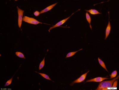
SH-SY5Y cell; 4% Paraformaldehyde-fixed; Triton X-100 at room temperature for 20 min; Blocking buffer (normal goat serum, SLC0005) at 37°C for 20 min; Antibody incubation with (PI 3 Kinase p85 alpha) monoclonal Antibody, Unconjugated (SLM33219M) 1:100, 90 minutes at 37°C; followed by a conjugated Goat Anti-Mouse IgG antibody at 37°C for 90 minutes, DAPI (blue, C02-04002) was used to stain the cell nuclei.
|