[IF=4.546] Kuankuan Xiong. et al. Low-Concentration T-2 Toxin Attenuates Pseudorabies Virus Replication in Porcine Kidney 15 Cells. Toxins. 2022 Feb;14(2):121 WB ; Pig.
[IF=4.411] Guodong Zhao. et al. Isolation and Characterization of Natural Nanoparticles in Naoluo Xintong Decoction and Their Brain Protection Research. Molecules. 2022 Jan;27(5):1511 WB ; Rat.
[IF=5.076] Chao Zhang. et al. Affibody Modified G-quadruplex DNA Micelles Incorporating Polymeric 5-Fluorodeoxyuridine for Targeted Delivery of Curcumin to Enhance Synergetic Therapy of HER2 Positive Gastric Cancer. Nanomaterials-Basel. 2022 Jan;12(4):696 WB ; Human.
[IF=3.252] Guangcong Peng. et al. Intranasal administration of DHED protects against exhaustive exercise-induced brain injury in rats. Brain Res. 2021 Dec;1772:147665 IF ; rat.
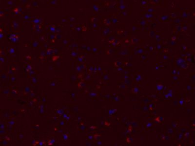
[IF=16.806] Min Qian. et al. Molybdenum Diphosphide Nanorods with Laser-Potentiated Peroxidase Catalytic/Mild-Photothermal Therapy of Oral Cancer. 2021 Oct 31 IHC ; Mouse.
[IF=2.795] Ying Feng. et al. LncRNA MALAT1 inhibits apoptosis of endometrial stromal cells through miR-126-5p-CREB1 axis by activating PI3K-AKT pathway. Mol Cell Biochem. 2020 Dec;475(1):185-194 WB ; Human.
[IF=3.416] Zhang F et al. Affibody‐Conjugated RALA Polymers Delivering Oligomeric 5‐Fluorodeoxyuridine for Targeted Therapy of HER2 Overexpressing Gastric Cancer. Macromol Biosci
. 2020 Jul;20(7):e2000083. WB ; Human.
[IF=3.067] Kang Z et al. Copper-induced apoptosis and autophagy through oxidative stress-mediated mitochondrial dysfunction in male germ cells. Toxicol In Vitro. 2019 Sep 3;61:104639. WB ; Mouse.
[IF=1.664] Wang Z et al. β-Bourbonene attenuates proliferation and induces apoptosis of prostate cancer cells.Oncol Lett. 2018 Oct;16(4):4519-4525. WB ; Human.
[IF=3.269] Leiming Sun. et al. Isoliquiritigenin attenuates acute renal injury through suppressing oxidative stress, fibrosis and JAK2/STAT3 pathway in streptozotocin-induced diabetic rats. Bioengineered. 2021;12(2):11188-1240 WB ; Rat.