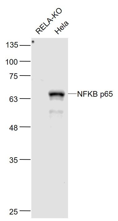
Sample:
RELA-KO(Human) Cell Lysate at 30 ug
Hela(Human) Cell Lysate at 30 ug
Primary: Anti-NFKB p65 (SLM33117M) at 1/1000 dilution
Secondary: IRDye800CW Goat Anti-Mouse IgG at 1/20000 dilution
Predicted band size: 65 kD
Observed band size: 65 kD
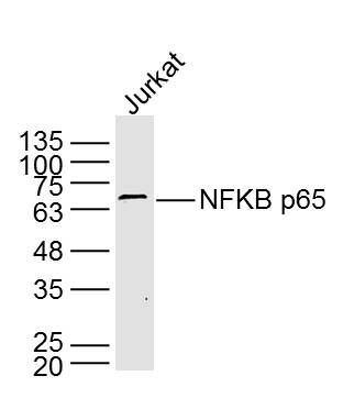
Sample: Jurkat Cell (Human) Lysate at 40 ug
Primary: Anti- NFKB p65 (SLM33117M) at 1/2 000 dilution
Secondary: IRDye800CW Goat Anti-Mouse IgG at 1/20000 dilution
Predicted band size: 61 kD
Observed band size: 65 kD
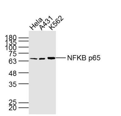
Sample:
Hela Cell (Human) Lysate at 40 ug
A431 Cell (Human) Lysate at 40 ug
K562 Cell (Human) Lysate at 40 ug
Primary: Anti- NFKB p65 (SLM33117M) at 1/1000 dilution
Secondary: IRDye800CW Goat Anti-Mouse IgG at 1/20000 dilution
Predicted band size: 61 kD
Observed band size: 65 kD
Sample:
HL60A549(Human) Cell Lysate at 30 ug
Primary: Anti-NFKB p65 (SLM33117M) at 1/1000 dilution
Secondary: IRDye800CW Goat Anti-MouseIgG at 1/20000 dilution
Predicted band size: 65 kD
Observed band size: 62 kD
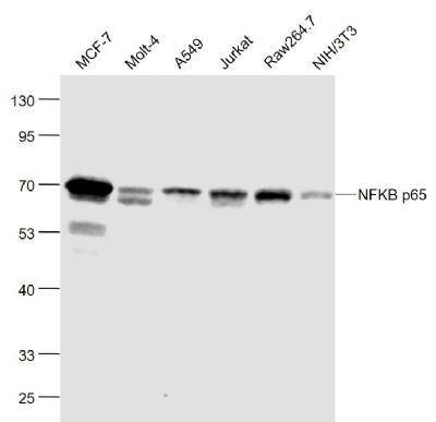
Sample:
MCF-7(Human) Cell Lysate at 30 ug
Molt-4(Human) Cell Lysate at 30 ug
A549(Human) Cell Lysate at 30 ug
Jurkat(Human) Cell Lysate at 30 ug
Raw264.7(Mouse) Cell Lysate at 30 ug
NIH/3T3(Mouse) Cell Lysate at 30 ug
Primary: Anti-NFKB p65 (SLM33117M) at 1/1000 dilution
Secondary: IRDye800CW Goat Anti-Mouse IgG at 1/20000 dilution
Predicted band size: 65 kD
Observed band size: 62 kD
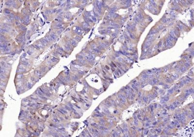
Paraformaldehyde-fixed, paraffin embedded (Rat colon); Antigen retrieval by boiling in sodium citrate buffer (pH6.0) for 15min; Block endogenous peroxidase by 3% hydrogen peroxide for 20 minutes; Blocking buffer (normal goat serum) at 37°C for 30min; Antibody incubation with (NFKB p65) Monoclonal Antibody, Unconjugated (SLM33117M) at 1:500 overnight at 4°C, followed by a conjugated secondary (sp-0023) for 20 minutes and DAB staining.
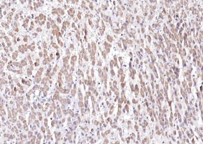
Paraformaldehyde-fixed, paraffin embedded (human liver); Antigen retrieval by boiling in sodium citrate buffer (pH6.0) for 15min; Block endogenous peroxidase by 3% hydrogen peroxide for 20 minutes; Blocking buffer (normal goat serum) at 37°C for 30min; Antibody incubation with (NFKB p65) Monoclonal Antibody, Unconjugated (ascites of SLM33117M 7G6) at 1:2000 overnight at 4°C, followed by operating according to SP Kit(Mouse) (sp-0024) instructions and DAB staining.
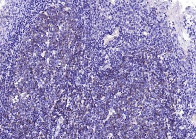
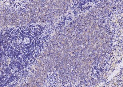
Paraformaldehyde-fixed, paraffin embedded (human tonsil); Antigen retrieval by boiling in sodium citrate buffer (pH6.0) for 15min; Block endogenous peroxidase by 3% hydrogen peroxide for 20 minutes; Blocking buffer (normal goat serum) at 37°C for 30min; Antibody incubation with (NFKB p65) Monoclonal Antibody, Unconjugated (ascites of SLM33117M 7G6) at 1:2000 overnight at 4°C, followed by operating according to SP Kit(Mouse) (sp-0024) instructions and DAB staining.
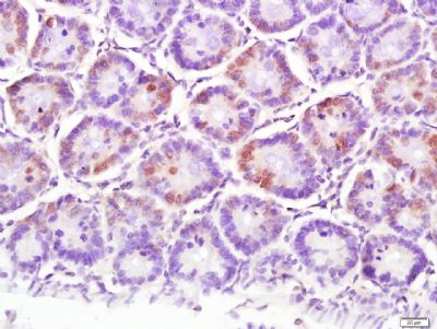
Paraformaldehyde-fixed, paraffin embedded (human colon carcinoma); Antigen retrieval by boiling in sodium citrate buffer (pH6.0) for 15min; Block endogenous peroxidase by 3% hydrogen peroxide for 20 minutes; Blocking buffer (normal goat serum) at 37°C for 30min; Antibody incubation with (NFKB p65) Monoclonal Antibody, Unconjugated (ascites of SLM33117M 7G6) at 1:2000 overnight at 4°C, followed by operating according to SP Kit(Mouse) (sp-0024) instructions and DAB staining.
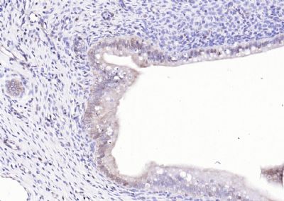
Paraformaldehyde-fixed, paraffin embedded (rat uterus); Antigen retrieval by boiling in sodium citrate buffer (pH6.0) for 15min; Block endogenous peroxidase by 3% hydrogen peroxide for 20 minutes; Blocking buffer (normal goat serum) at 37°C for 30min; Antibody incubation with (NFKB p65) Monoclonal Antibody, Unconjugated (ascites of SLM33117M 7G6) at 1:2000 overnight at 4°C, followed by operating according to SP Kit(Mouse) (sp-0024) instructions and DAB staining.
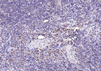
Paraformaldehyde-fixed, paraffin embedded (rat spleen); Antigen retrieval by boiling in sodium citrate buffer (pH6.0) for 15min; Block endogenous peroxidase by 3% hydrogen peroxide for 20 minutes; Blocking buffer (normal goat serum) at 37°C for 30min; Antibody incubation with (NFKB p65) Monoclonal Antibody, Unconjugated (ascites of SLM33117M 7G6) at 1:2000 overnight at 4°C, followed by operating according to SP Kit(Mouse) (sp-0024) instructions and DAB staining.
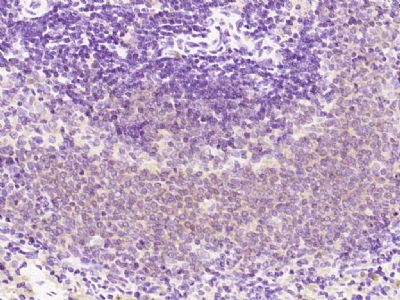
Paraformaldehyde-fixed, paraffin embedded (mouse spleen); Antigen retrieval by boiling in sodium citrate buffer (pH6.0) for 15min; Block endogenous peroxidase by 3% hydrogen peroxide for 20 minutes; Blocking buffer (normal goat serum) at 37°C for 30min; Antibody incubation with (NFKB p65) Monoclonal Antibody, Unconjugated (ascites of SLM33117M 7G6) at 1:2000 overnight at 4°C, followed by operating according to SP Kit(Mouse) (sp-0024) instructions and DAB staining.
Paraformaldehyde-fixed, paraffin embedded (rat spleen); Antigen retrieval by boiling in sodium citrate buffer (pH6.0) for 15min; Block endogenous peroxidase by 3% hydrogen peroxide for 20 minutes; Blocking buffer (normal goat serum) at 37°C for 30min; Antibody incubation with (NFKB p65) Monoclonal Antibody, Unconjugated (SLM33117M) at 1:200 overnight at 4°C, followed by operating according to SP Kit(Mouse)(sp-0024) instructionsand DAB staining.
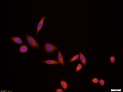
Tissue/cell:MCF7 cell; 4% Paraformaldehyde-fixed; Triton X-100 at room temperature for 20 min; Blocking buffer (normal goat serum,SLC0005) at 37°C for 20 min; Antibody incubation with (NFKB p65) monoclonal Antibody, Unconjugated (SLM33117M)1:100, 90 minutes at 37°C; followed by a CY3 conjugated Goat Anti-Mouse IgG antibody at 37°C for 90 minutes, DAPI (blue, C02-04002) was used to stain the cell nuclei.
|