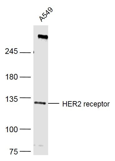
Sample:
A549(Human) Cell Lysate at 40 ug
Primary: Anti-HER2 receptor (SLM2156R) at 1/500 dilution
Secondary: IRDye800CW Goat Anti-Mouse IgG at 1/20000 dilution
Predicted band size: 138 kD
Observed band size: 134 kD
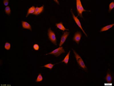
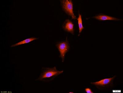
Tissue/cell:A549 cell; 4% Paraformaldehyde-fixed; Triton X-100 at room temperature for 20 min; Blocking buffer (normal goat serum,SLC0005) at 37°C for 20 min; Antibody incubation with (HER2 receptor) monoclonal Antibody, Unconjugated (SLM2156M) 1:100, 90 minutes at 37°C; followed by a CY3 conjugated Goat Anti-Mouse IgG antibody at 37°C for 90 minutes, DAPI (blue, C02-04002) was used to stain the cell nuclei.
Tissue/cell:A549 cell; 4% Paraformaldehyde-fixed; Triton X-100 at room temperature for 20 min; Blocking buffer (normal goat serum,SLC0005) at 37°C for 20 min; Antibody incubation with (HER2 receptor) monoclonal Antibody, Unconjugated (SLM2156M) 1:100, 90 minutes at 37°C; followed by a CY3 conjugated Goat Anti-Mouse IgG antibody at 37°C for 90 minutes, DAPI (blue, C02-04002) was used to stain the cell nuclei.
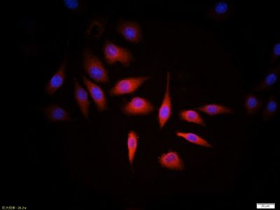
Tissue/cell:MCF7 cell; 4% Paraformaldehyde-fixed; Triton X-100 at room temperature for 20 min; Blocking buffer (normal goat serum, SLC0005) at 37°C for 20 min; Antibody incubation with (HER2 receptor) monoclonal Antibody, Unconjugated (SLM2156M) 1:100, 90 minutes at 37°C; followed by a CY3 conjugated Goat Anti-Mouse IgG antibody at 37°C for 90 minutes, DAPI (blue, C02-04002) was used to stain the cell nuclei.
|