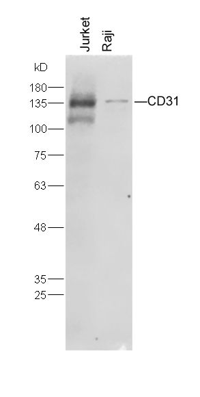
Protein:
Jurkat(human) cell lysates at 20ug;
Raji(human) cell lysates at 20ug;
Primary: Anti-CD31 (SLM10825M, 1A5) at 1:300;
Secondary: IRDye800CW Conjugated Goat (polyclonal) Anti-Mouse IgG(H+L) (SL40296G-IRDye8) at 1: 20000;
Predicted band size : 78kD
Observed band size : 140kD
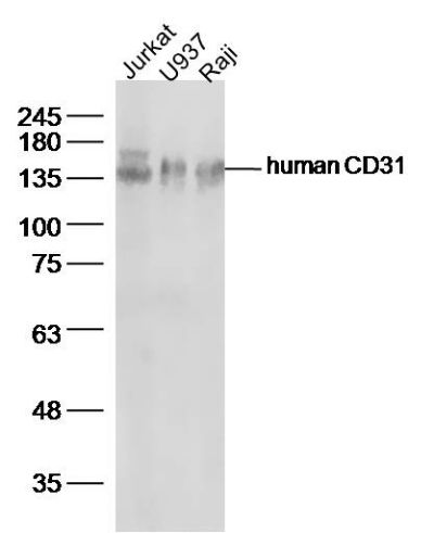
Sample:
Jurkat(human)cell Lysate at 30 ug
U937(human)cell Lysate at 40 ug
Raji(human)cell Lysate at 40 ug
Primary: Anti-human CD31(SLM10825MR) at 1/300 dilution
Secondary: IRDye800CW Goat Anti-Rabbit IgG at 1/20000 dilution
Predicted band size: 78 kD
Observed band size: 140 kD
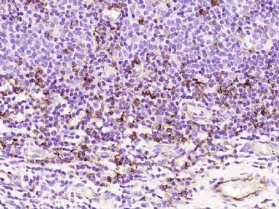
Paraformaldehyde-fixed, paraffin embedded (human tonsil); Antigen retrieval by boiling in sodium citrate buffer (pH6.0) for 15min; Block endogenous peroxidase by 3% hydrogen peroxide for 20 minutes; Blocking buffer (normal goat serum) at 37°C for 30min; Antibody incubation with (CD31) Monoclonal Antibody, Unconjugated (ascites of SLM10825M 3B3) at 1:2000 overnight at 4°C, followed by operating according to SP Kit(Mouse) (sp-0024) instructions and DAB staining.
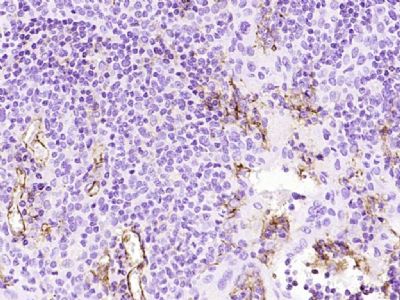
Paraformaldehyde-fixed, paraffin embedded (human tonsil); Antigen retrieval by boiling in sodium citrate buffer (pH6.0) for 15min; Block endogenous peroxidase by 3% hydrogen peroxide for 20 minutes; Blocking buffer (normal goat serum) at 37°C for 30min; Antibody incubation with (CD31) Monoclonal Antibody, Unconjugated (ascites of SLM10825M 2D4) at 1:2000 overnight at 4°C, followed by operating according to SP Kit(Mouse) (sp-0024) instructions and DAB staining.
|