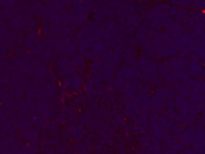
Paraformaldehyde-fixed, paraffin embedded (Rat thymus); Antigen retrieval by boiling in sodium citrate buffer (pH6.0) for 15min; Block endogenous peroxidase by 3% hydrogen peroxide for 20 minutes; Blocking buffer (normal goat serum) at 37°C for 30min; Antibody incubation with (Collagen I) Monoclonal Antibody, Unconjugated (SLM33400M) at 1:400 overnight at 4°C, followed by a conjugated Goat Anti-Mouse IgG antibody (SL0296G-CY3) for 90 minutes, and DAPI for nuclei staining.
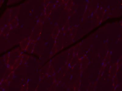
Paraformaldehyde-fixed, paraffin embedded (Rat skeletal muscle); Antigen retrieval by boiling in sodium citrate buffer (pH6.0) for 15min; Block endogenous peroxidase by 3% hydrogen peroxide for 20 minutes; Blocking buffer (normal goat serum) at 37°C for 30min; Antibody incubation with (Collagen I) Monoclonal Antibody, Unconjugated (SLM33400M) at 1:400 overnight at 4°C, followed by a conjugated Goat Anti-Mouse IgG antibody (SL0296G-CY3) for 90 minutes, and DAPI for nuclei staining.