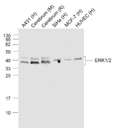
Sample:
Lane 1: A431 (Human) Cell Lysate at 30 ug
Lane 2: Cerebrum (Mouse) Lysate at 40 ug
Lane 3: Cerebrum (Rat) Lysate at 40 ug
Lane 4: SiHa (Human) Cell Lysate at 30 ug
Lane 5: MCF-7 (Human) Cell Lysate at 30 ug
Lane 6: HUVEC (Human) Cell Lysate at 30 ug
Primary: Anti-ERK1/2 (SLM33337M) at 1/1000 dilution
Secondary: IRDye800CW Goat Anti-Mouse IgG at 1/20000 dilution
Predicted band size: 44/42 kD
Observed band size: 40/42 kD
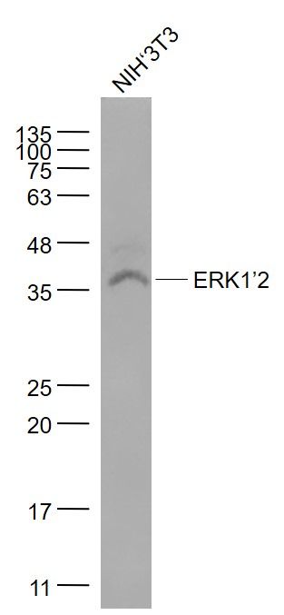
Sample:
NIH/3T3(Mouse) Cell Lysate at 30 ug
Primary: Anti- ERK1/2 (SLM33337M) at 1/1000 dilution
Secondary: IRDye800CW Goat Anti-Rabbit IgG at 1/20000 dilution
Predicted band size: 43 kD
Observed band size: 38 kD
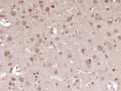
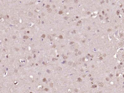
Paraformaldehyde-fixed, paraffin embedded (mouse brain); Antigen retrieval by boiling in sodium citrate buffer (pH6.0) for 15min; Block endogenous peroxidase by 3% hydrogen peroxide for 20 minutes; Blocking buffer (normal goat serum) at 37°C for 30min; Antibody incubation with (ERK1) Monoclonal Antibody, Unconjugated (ascites of SLM33337M) at 1:2000 overnight at 4°C, followed by operating according to SP Kit(Mouse) (sp-0024) instructions and DAB staining.
Paraformaldehyde-fixed, paraffin embedded (mouse brain); Antigen retrieval by boiling in sodium citrate buffer (pH6.0) for 15min; Block endogenous peroxidase by 3% hydrogen peroxide for 20 minutes; Blocking buffer (normal goat serum) at 37°C for 30min; Antibody incubation with (ERK1) Monoclonal Antibody, Unconjugated (ascites of SLM33337M) at 1:2000 overnight at 4°C, followed by operating according to SP Kit(Mouse) (sp-0024) instructions and DAB staining.
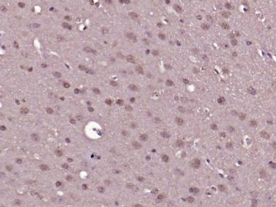
Paraformaldehyde-fixed, paraffin embedded (rat brain); Antigen retrieval by boiling in sodium citrate buffer (pH6.0) for 15min; Block endogenous peroxidase by 3% hydrogen peroxide for 20 minutes; Blocking buffer (normal goat serum) at 37°C for 30min; Antibody incubation with (ERK1) Monoclonal Antibody, Unconjugated (ascites of SLM33337M) at 1:2000 overnight at 4°C, followed by operating according to SP Kit(Mouse) (sp-0024) instructions and DAB staining.
|